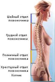
Spine is the axis of our body. It is also called the spinal column, which displays it appearance... It connects directly to almost all parts of the body: the skull, upper limbs, chest, pelvis, and through it with the lower limbs. But it consists not only of the bone part, because it is a highly organized structure.
Spinal nerve connections to the vertebrae
The first cervical nerve arises between the skull and the atlas, and the cervical nerves C2 to 7 continue to leave the spinal canal above the corresponding numbered vertebra.
Regional characteristics of the vertebrae
The vertebrae of each region have specific characteristics, which are now described.The seven vertebrae of the neck are characterized by a foramen in each transverse process known as the transversary foramen. The atlas, which has neither body nor spinous, consists of two lateral masses connected by a short anterior and a longer posterior arch. Each lateral mass represents the superior and inferior faces, for the occipital condyle of the skull and for the axis, respectively. The anterior arch represents the anterior tubercle in the front and the facet in the back.
The spine is an important part of the human body. Thanks to him, our posture is even, we can stand and walk, and live a full and active life.
The main structure of the spinal column is the vertebra. There are from 32 to 34 such structures in the body, depending on the physiological characteristics. Our spine is mobile, due to which the person himself is mobile. This is explained by the fact that there are special formations between our vertebrae, through which our vertebrae do not rub against each other. These formations include intervertebral (intervertebral) discs, ligamentous apparatus of the spine, facet (vertebral) joints.
The posterior arch is grooved higher for the vertebral artery and a small C1 nerve on each side and represents the posterior posterior tubercle. The axis is characterized by teeth that protrude upward from the body and articulate with the front arch of the satin. Lairs are attached to the occipital bone and are bounded behind transverse ligament atlas. It is often claimed that the lairs represent the body of an atlas, but this is doubtful.
Remaining cervical vertebrae
The lower cervical vertebrae, C2-6, are typical and are short, bivalve spinous processes. The organs are small and oval in shape. On the superolateral borders of the vertebral bodies there are lips with excellent projection. They closely correspond to the indentations in the lateral border of the lower aspect of the vertebral body above. Each transverse process punctured by the skeleton of the carcass ends laterally in the anterior and posterior tubercles, which are connected by an "intertubular plate" or bar. The rods are cut with ventral primary rams spinal nerves that pass behind the transversal.
Normally, in an adult, some vertebrae grow together into separate bones, and, accordingly, there is no normal mobility between these vertebrae. These are the vertebrae of the sacral and coccygeal spine. They grow together to form solid bones: sacral and coccygeal bones.
As already mentioned, the spine, based on physiological characteristics, has from 32 to 34 vertebrae and it is customary to divide it into several sections, consisting of a certain number of such vertebrae. Each department and each vertebra has its own designations. For greater convenience, each section has its own letter, based on the Latin name of the spine section:
Thoracic vertebrae
It should be noted that all vertebrae are dorsal, although only 12 are thoracic. The 12 vertebrae of the chest bear the ribs. Defacts for the heads of the ribs were found lower and lower at the junction of the body and the leg. The spinous processes are long, thin and oblique: their tips lie opposite the lower body or even at the level of the intervertebral disc below the lower body. The hunchback back is called kyphosis.
Lumbar vertebrae
The five vertebrae between the thorax and the sacrum are large and do not represent either transversals or articular edges.The body is renal wall, and the legs and plates are short and thick. The mamillary process develops backward from the superior articular process. The transverse process corresponding to an edge is long and thin, and an auxiliary process can be projected below its root. The spinous processes are quadrangular and project horizontally backward. The fifth lumbar vertebra, usually the largest vertebra, is mainly responsible for the lumbosacral angle.
- cervical spine - cervical (pars cervicalis) - C;
- thoracic spine - thoracic (pars thoracalis) - Th or T, can also be denoted - D;
- lumbar spine - lumbar (pars lumbalis) - L;
- sacral spine - sacrum bone (os sacrum) - S;
- coccygeal spine - coccygeal bone (os coccygis) - Co.
The numbering of the vertebrae in each of its sections starts from the top, increasing in number downward:
Sacrum
An excessive "notch" in the back is called lordosis. The five vertebrae are fused in the adult to form the sacrum, which can be felt below the "lesser back". It has a roughly triangular appearance with a pelvic and dorsal surface, a lateral mass on each side, and a base and apex. The spinal nerve anesthetic can be administered extradurally through the sacral break. The lateral part or mass of the sacrum, lateral to the sacral foramini, consists of fused transverse processes. Its excellent surface is often referred to as scarlet.
- there are 7 vertebrae in the cervical spine, respectively - C1 - C7; the occipital bone of the skull is conventionally considered the zero cervical vertebra, respectively - C0;
- in the chest - 12, respectively - Th1 - Th12(or T1 - T12, or D1 - D12);
- in the lumbar - 5, respectively - L1 - L5;
- in the sacred - 5, respectively - S1 - S5;
- and in the coccygeal from 3 to 5, respectively - Co1 - Co5.
Features of the vertebrae cervical
The 1st, 2nd and 7th cervical vertebrae differ in structure from the rest of the vertebrae, and have their own names:
The supolateral portion of the lateral mass is the surface of the auricular portion for articulation with the hip bone. The surface is limited by the area for the interosseous ligaments. The base formed by the obverse of the first sacral vertebra is the prominent anterior margin, designated the promontory.
Articular articular processes are combined with the fifth lumbar vertebra. The sacral canal extends from the base to the sacral hiatus. The apex of the sacrum can be fused with the coccyx. The vertebrae below the sacrum intertwine alternately in adults to form a coccyx that resembles a miniature sacrum in shape.
- C1 is called Atlas - in Latin Atlas;
- C2 is called the Axial vertebra or Axis or Epistrophy - in Latin Axis;
- C7 is called Protruding vertebra - in Latin Vertebra Prominens.
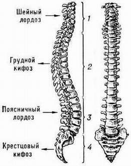
Not everyone knows, but in fact, our spine is not perfectly straight, because it has four physiological bends in the sagittal plane (side view), due to which the spinal column is absorbed, not damaged and due to which it can distribute the load of the human body weight. In addition, it protects our brain from mechanical damage (concussions).
Development of the vertebrae
Vertebrae develop in the mesenchyme and cartilage during the actual embryonic period, and most begin to ossify during fetal life. Typically, the spine at birth shows three ossiphic regions, one for the center and one for each half of the neural arch. During puberty, ossiphic centers appear on the fields of the upper and lower surfaces of the body and at the tips of various processes. One of the reasons for the lateral curvature is the refusal to develop half of the vertebra.
The malfunction of the fusion of the halves of one or more neural arches is called spina bifida. Violations only in the form of meninges or spinal cord and meninges can protrude through the defect. This is quite common in the sacrum. The ocular processes of the vertebrae are palpable in the median sulcus of the back. External occipital dislocation is noticeable in adults. In the thoracic region, the spinous process of each vertebra extends to the level of at least the vertebral body below.
There are two types of such bends: lordosis and kyphosis. Lordosis is the anterior bending of the spinal column (ventrally - i.e. to the side internal organs in relation to the midline of the body), and kyphosis is its bending posteriorly (dorsally - i.e. towards the spine in relation to the midline of the body). We have two lordosis and two kyphosis. These curves are:
- Cervical lordosis - in the cervical (cervical) region, the vertebral column bends slightly forward (ventrally);
- Thoracic kyphosis - the bend of its thoracic (thoracic) section backward (dorsally);
- Lumbar lordosis is an anterior (ventral) bend of the lumbar (lumbar) section;
- Sacral kyphosis is the bending of its sacral (sacral) section backward (dorsally).
Structure, functions of the vertebrae
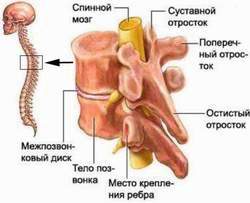
Vertebra(in Latin " vertebra") consists of several parts:
The posterior superior iliac spine is usually marked with a cutaneous depression. This classic study of the entire skeleton is available in a later edition in German. It is an interesting procedure in pain medicine that uses specialized devices to alter nerve conduction. This relief pain relief method can be used for patients suffering from back pain, neck pain, muscle pain, or other multiple pain syndromes. The probe is inserted into the needle and uses pain to force controlled heat into the affected nerve.
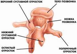
- the vertebral body (its most massive part), which can be both round and kidney-shaped;
- vertebral arch, playing the role of a closure of the vertebral foramen, in which is located spinal cord;
- 7 processes: (a) paired upper and lower articular processes (a total of four for each vertebra), due to which the vertebrae located above and below are combined with each other, (b) paired transverse processes (only two for each vertebra) and ( c) one unpaired spinous process (directed posteriorly / dorsally / from the arch).
Due to the presence of paired articular processes in the vertebrae above and below, each vertebra can be connected to another vertebra by vertebral (facet, facet) joints. In addition to these joints, the vertebrae are connected by cartilaginous formations: (a) intervertebral (or intervertebral) discs located between the vertebral bodies, (b) long and short ligaments of the spinal column, located both on the anterior and posterior surfaces of the vertebrae and its processes, and muscles of three types: (a) rotator muscles, (b) multifidus muscles, and (c) transverse muscles.
Radiofrequency ablation does not damage the nerves, but it is used to neutralize painful nerves. The anatomical and physiological function of the spine is the key to assessing a person associated with spinal pain problems. The bone column is positioned so that individual vertebrae can provide a flexible structural support, thus protecting the spinal cord. The borders of the spine are a mobile connection that connects one vertebra to another. The veneers are innervated by the medial nerve of the ramus, which provides sensation to the joints.
Thanks to the discs, ligaments, joints and muscles, our vertebrae are mobile. An important role of the ligaments is that they prevent the vertebrae from moving too much and go beyond their anatomical boundaries.
In many ways, how mobile the spinal column is depends on the strength and training of our muscles that support it, these are the muscles: dorsal, chest, shoulder, cervical, femoral, and also abdominal muscles. Thanks to these muscles, we control the movements of our spine, they are soft and full. These muscles perfectly interact with each other, and if any muscle goes beyond such a harmonious state, this affects the entire motor activity of the spinal column. In addition to the fact that his functional state is disturbed, a feeling of discomfort and soreness in the back may also appear.
Joints can become arthritic or affected, causing severe pain with activity. The medial branches are branches of small nerves that communicate pain to the brain caused by the articular edges in the spine. The nerve is located in the joints of the transverse process and the articular articular joint. Radiofrequency ablation of the medial branches is minimally invasive for the patient and reduces back pain by interrupting the nerve supply to the articular edges of the neck or back.
A ganglion is a collection of nerves. The fragmented palatine ganglion provides innervation to the face and head. A recent article shows that radiofrequency can be treated in the ganglion for chronic headaches or other conditions that cause unusual facial pain.
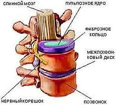
The intervertebral discs located between the vertebrae of the spinal column of the neck, chest and lower back are of great importance (with the exception of the first and second cervical vertebrae). The composition of such a disc includes a gelatinous pulpous nucleus and a multilayer annulus fibrosus. The disc is so elastic in structure that it can change its shape. Thanks to this feature, he is able to control the degree of movement of the vertebrae and cushion them for a softer and more non-traumatic movement.
Radiofrequency ablation is a process that creates nerve injury caused by localized heat. When trauma is created on a nerve, pain signals are interrupted and the perception of pain that the brain produces is diminished. This process is performed in an external patient environment. This procedure is performed using local anesthesia in combination with a sedative serum when needed. Before starting the process, the doctor will begin disinfecting the skin where the injection will be placed and then injecting local anesthesia to numb the skin.
Another needle is placed through the sleeping tissue and the process is performed using guided X-rays. When the needle is in the correct position, the electrode is inserted into the center of the needle. Stimulation starts with sensory stimulation and then motor stimulation. When is checked correct position local anesthesia and sometimes steroid medicine are administered. Thermal heat is generated, which allows the fibers surrounded by pain to be destroyed, thereby relieving pain. This method is less inconvenient and usually has a moderate pulsation sensation.
Outside, the vertebra is covered with a dense substance, fencing it off from nearby tissues and structures, damage, inside it is filled with a softer spongy substance. The vertebra is strong enough also due to the fact that this very spongy substance creates special crossbeams. The compact substance that covers the outside of the vertebra is formed from lamellar bone tissue, which gives it firmness and endurance while a person performs various kinds of physical activity, such as walking. Another important content of the vertebra is red Bone marrow, thanks to which red blood cells are formed in our body. It should be said that the red bone marrow is found to a greater extent in the pelvic bones, to a lesser extent in the tubular bones of the skeleton and, to a lesser extent, in the vertebral bodies.
After a period of mild recovery, you can return home after treatment. You may have mild discomfort when local anesthetic ends in your body. In a recent clinical study for patients receiving radiofrequency therapy, 21% had complete pain relief and 65% had moderate to moderate pain relief. Most of the respondents reported a decrease in the use of pain medications. None of the patients developed significant infections, bleeding, bruising, or numbness from complications of their therapy.
Patients can experience great relief from a minimally invasive process that does not require hospitalization. As with any medical care, there are risks and possible complications. However, there is rarely a complication. Possible complications that may occur include: Bleeding, infection, symptoms of increased pain, discomfort during injection, and rarely nerve damage. The relief received from radio frequencies is between three and six months.
Despite the fact that the structure of all vertebrae is, in principle, identical, they still differ in the morphology of their bodies and processes, that is, the vertebrae of each section of the spinal column have their own differences. The most massive and largest bodies and processes have vertebrae located in the lumbar (lumbar) region. This can be explained. The fact is that the greatest load falls on the lumbar spine, which "forced" nature to make these vertebrae the strongest and most enduring. The vertebral bodies of the cervical (cervical) spine are small and not so massive, because their main task is to hold the head, which is why these vertebrae are quite tender and easily injured, therefore, you need to be careful with the neck, especially when playing sports.
Radiofrequency treatments are very safe, wearable methods of choice for treating a variety of causes of chronic pain. If your pain responds well to treatment, re-treatment is recommended. A number of 100 patients with trigeminal neuroglia were treated with radiofrequency thermocoagulation. In another study, thirty-nine patients were assessed with trigeminal neuralgia and 3% received satisfactory pain relief. The American Society of Interval Pain Physicians has developed a practice guideline based on large evidence for the management of chronic back pain to guide clinics.
It is worth mentioning separately about the vertebrae that form the thoracic (thoracic) spine, because they create the chest together with the sternum and ribs. The vertebrae have transverse processes, to which the ribs are attached, so it is wrong to consider the spine and ribs as one whole, because between them there are special articular structures that provide the chest with its mobility. It is the movable ribs, as well as the ability to move between the vertebrae and ribs, that help the chest not to create an obstacle to the implementation of the most important function - breathing, namely, to exhale and inhale. By itself, the thoracic spine is not as mobile as the lower back or neck, and the space between the thoracic vertebrae is somewhat less than between the vertebrae of the lumbar spine.
For the treatment of middle branches, they state that repeated injections at regular intervals provide lasting pain relief. For mediacentral radiofrequency neurotomy, short-term pain relief was defined with a time of less than three months and long-term relief of 3 or more months. If you suffer from back, neck, face, or other painful conditions, see a pain specialist in Arizona to see if you can benefit from this cutting edge technology.
We are delighted to offer this service in the Phoenix area. The vertebral column is part of the body structure of vertebrates. characterizes the animals of the vertebrate group, and they constitute the subflume of chordates, agates, fish, amphibians, reptiles, birds and mammals, as explained. They are characterized by the presence of a segmented vertebral column and skull. protects the brain. In humans, the spine or spine almost always forms. on 33 and ultimately 32 or 34 vertebrae, which are connected by joints that. are of two types: a larger one with interpolation of intervertebral discs in. the anterior region between each vertebra and two smaller vertebrae, for a double pair. rear border edges, two sides up and two opposite each other. down, forming on each side, behind, in the vertebra, a joint. chamfer.
These are holes that form between a pair of nearby (adjacent) vertebrae. The corresponding roots pass through these holes from the spinal cord, as well as vessels: arteries and veins. From receptors located in the thickness of the skin layer, as well as from the fibrous layers of connective tissue, nerve impulses through the fibers of the spinal roots go directly to the spinal cord. There are other nerve fibers whose task is to transmit signals and impulses from the spinal cord to the muscles, causing them to contract under the command of both the spinal cord and the brain. Each level of the spinal cord is responsible for the innervation of specific parts of the body. So, in "a few words" we can say that the roots of the cervical spine innervate upper limbs, roots lumbar- the lower limbs, and the roots of the thoracic region - the body (trunk).
The opinion that bones do not undergo changes is erroneous, but this is not so, because throughout their life, bone cells are constantly renewed: some of them die, and new ones are formed to replace them. Due to the fact that we are in constant motion, the formation of new bone tissue cells is stimulated. From this it is worth concluding that the more active a person leads, the younger his skeleton and spine, including! Accordingly, the less a person moves, the faster his spinal column ages.
Forced immobility of the spine
It is unpleasant that if a person is forced to move due to injury, illness or for other reasons for a long time, then bone gradually thinns, as a result of which the bones of the skeleton (including the spine) gradually soften. In such cases, in order to prevent the softening of the bones of the skeleton, the specialists of our Clinic recommend to undergo a course of physiotherapy (with the help of an electrical myostimulation apparatus) and therapeutic massage, which can be done in our Clinic.
Vertebral-motor segments (spinal PDS)
The term " spinal motion segment" or Vertebral column(or PDS of the vertebral column) means a section of the spinal column, consisting of a pair of adjacent (adjacent top-bottom) vertebrae.
The spinal motion segment includes all those structures of the spinal column that are located at the appropriate level. These are structures such as the vertebrae with their joints and ligamentous apparatus, the intervertebral disc and even the muscles surrounding the spine (paravertebral). Each such vertebral-motor segment has a pair of foraminal (intervertebral) openings, which serve as a receptacle for the roots of the spinal cord, as well as arteries and veins.
There are 24 such spinal motion segments in the spinal column. These are seven cervical SMS, twelve thoracic SMS and five lumbar SMS, with the last such segment forming L5 (fifth lumbar vertebra) and S1 (first sacral vertebra).
Disorders in the work of the PDS "s of the spine
With various dysfunctions of the spinal column, reversible so-called "blocks" are formed in the spinal motion segments, in which the facet (vertebral) joints are blocked (stop moving in the physiological volume), and the vertebrae are displaced from their physiologically correct positions, which causes a feeling of discomfort and soreness not only in a certain area of the back, but also, possibly, in the limbs, as well as other symptoms. In this case, the tasks of the chiropractor will be:
- elimination of blocks (dysfunctions) of the PDS of the spine;
- normalization of the range of motion in the joints of the spine;
- normalization of the tone of the paravertebral muscles;
- elimination of the causes of pain and other symptoms;
- consolidation of the achieved results for a long time.
To solve the above problems, our Clinic has qualified personnel and the necessary modern equipment.
The vertebrae are called vertebre in Latin, and the science that studies the spine and its pain is called vertebrology. Sometimes in the diagnosis you can find the word vertebral or vertebral, which means "descended from the spine."
In general terms, all vertebrae have a similar structure, however, depending on which department the vertebrae belong to and what predominant loads they experience, the vertebrae acquire certain characteristic outlines.
Each vertebra has a body - the most massive part of the vertebra, with which it rests on the underlying and on which the overlying vertebrae rest. This part of the vertebra has a great ability to withstand compressive force, has a cylindrical shape and gradually increases in diameter from the cervical vertebrae to the lower back.
In addition to the body, each vertebra of any department has an arc in the back that connects to the body with the help of legs. The body and arch define the vertebral foramen. The vertebral foramen of all vertebrae form the vertebral canal, which contains the spinal cord with membranes, vessels and nerves. The processes extend from the arch of the vertebra in different directions: an unpaired spinous process is directed posteriorly, paired transverse processes are located on the sides, and paired upper and lower articular processes are located above and below the arch.
In the area of the legs of the arc, there are the upper and lower incisions, which, when the vertebrae overlap each other, form intervertebral foramen for the exit of the vessels and nerves from the spinal canal.
The structure of the cervical vertebrae is slightly different from all the others. The first of them - the atlas - does not have a body, but instead an anterior arch with lateral masses is formed. Second cervical vertebra- the axial (epistrophy) also has a very peculiar structure. Its body is connected to the Atlantean by the odontoid process. Both vertebrae constitute a unique mechanism due to which the movement of the head around the vertical axis and its tilt is carried out.
The transverse processes of all cervical vertebrae have holes (they are absent in other vertebrae), the imposition of the holes of one vertebra on the holes of another leads to the formation of a bony canal for the passage of vertebral arteries and nerves. Articular processes that take part in the formation of facet joints protrude above and below the arch. The articular surfaces on these processes are located in the horizontal plane. This contributes to the great mobility of the vertebrae of the cervical spine in the horizontal plane relative to each other (i.e. allows you to achieve a large angle of twisting of the cervical spine) - all this contributes to high mobility of the head. This was a very important achievement in the process of evolution, which made it possible to maintain a large angle of the horizon under visual control with minimal body mobility. However, this also provided a greater vulnerability of the cervical spine due to the relatively low mass and strength of the cervical vertebrae and their great mobility. The seventh cervical vertebra has the most prominent and prominent spinous process, which is a very convenient anatomical landmark when examining the spine.
The thoracic vertebrae also have their own structural features. This is primarily due to the presence of costal fossae on the bodies and transverse processes of the thoracic vertebrae for articulation with the heads and tubercles of the ribs. The spinous processes of the thoracic vertebrae are lowered down and overlap each other in a tile-like manner. On the articular processes of the thoracic vertebrae, the articular surfaces are projected in the frontal plane.
The lumbar vertebrae are much larger than other vertebrae, and the articular surfaces of their articular processes are located in the sagittal plane. These vertebrae carry the greatest load in the spinal column.
The sacrum has the shape of a pyramid with the base up and the top down. Its pelvic surface is concave, its posterior surface is convex. Its greatest bend is located in the region of the III vertebra. The sacrum consists of five fused sacral vertebrae. The fusion process begins at the age of 16 and ends completely by the age of 25. On the anterior and posterior surfaces of the sacrum, there are four sacral foramina through which the sacral nerves and vessels pass. There are 5 sacral ridges along the posterior convex surface of the sacrum. On the lateral masses of the sacrum, the articular surface is located - the junction with the pelvic bones, which is located at the level of the I and II sacral vertebrae, is covered with cartilage and is called the ear-shaped surface. Behind this surface is the tuberosity of the sacrum, to which ligaments are attached that strengthen the sacroiliac joint.
The base of the sacrum, connecting with the lower surface of the lumbar vertebra, forms a protrusion into the pelvic cavity, which is called the cape. The cape is important in determining the longitudinal dimensions of the pelvis and is important from the point of view of the shape of the spinal column. The sacrum is one of the most distinct parts of the male and female skeleton. In women, the sacrum is much wider, shorter and less curved than in men. This is due to the rearing of the child and childbirth: a woman needs a larger volume of the pelvic space and a smoother path for the baby to exit for a normal pregnancy and childbirth process.
The coccygeal vertebrae are located downward from the sacrum, its very top (there are usually four, sometimes five). Only the first coccygeal vertebra still retains traces of the general structure of the vertebrae, and the latter are in the form of balls. The coccygeal vertebrae often merge into one coccygeal bone - the coccyx. The muscles and fascia of the perineum are attached to the tailbone. In women, the coccyx is more mobile and during pregnancy it can be displaced (bent) posteriorly - to ensure the process of childbirth.