Content
First aid for damage internal organs...........................................................................................................
Conclusion………………….......................................... .................................
Conclusions................................................. .................................................. ...........
List of sources used ... ... ............................................. ........
Introduction
Children are very mobile and restless creatures. Showing curiosity and actively exploring the world around them, they often become victims of domestic trauma. Older children - can knock over a cup of hot tea, stick their fingers off the swing. It is not uncommon for schoolchildren to get injured while cycling and rollerblading.
Adults must know where the child is in danger, and do everything possible to prevent it. Alas, it is impossible to completely protect the child from injuries, and sooner or later children get bruises and bumps, burn themselves, break limbs, etc. The main task of adults in such a situation is not to panic, quickly call a doctor, and competently provide first aid, this will not only alleviate the child's condition, but also accelerate his subsequent recovery.
Relevance:
Purpose of the study: We will get acquainted with the main types of injuries and learn how to provide first aid for injuries.
Research objectives:
to repeat and consolidate the knowledge of students about the types of domestic injuries;
teach first aid (self-help) for various types of injuries; introduce the standard tools to the first medical care;
remind of precautions; develop joint action skills;
to educate a respectful attitude towards their health, the health of the people around them.
Object of study: is first aid.
Subject of study: rendering the first first aid victims to maintain and reduce damage to health until the arrival of an ambulance.
1 ... First aid for injuries
Trauma concept. Anatomical or functional disorders of tissues and organs resulting from the damaging effect of external factors on the human body are called trauma, or damage. Impacts can be mechanical (impact, squeezing, stretching), physical (exposure to heat and cold, electricity, radioactive radiation), chemical (due to the action of acids, alkalis, poisons), mental (fear, fear). The severity of damage depends on the severity and time of exposure to these factors.
Most often, damage is caused by mechanical influences, that is, by the direct action of mechanical force (impact, squeezing, stretching) on the tissues of the body. Mechanical damage can be closed and open. Closed damages are those in which there is no violation of the integrity skin and mucous membranes. These include bruises, sprains, subcutaneous tears soft tissues (muscles, tendons, blood vessels, nerves). Open injuries - damage to organs and tissues, accompanied by a violation of the integrity of the skin or mucous membranes (wounds, open bone fractures).
Injuries can also be thermal, chemical, electrical trauma, mental trauma, barotrauma, arising mainly after an explosion (due to the blast wave, the middle ear is damaged, the tympanic membrane is ruptured, this is accompanied by cranial manifestations - concussion, brain contusion).
Injuries resulting from a one-time, sudden strong effect on body tissues are called acute trauma, and those arising from repeated and constant effects of low force, unable to cause injury with a single action, are called chronic trauma. Chronic trauma includes most occupational diseases (flat feet in persons engaged in heavy physical labor, tendovaginitis in typists, chills in laundresses, eczema and ulcers on the hands of radiologists, etc.). Any trauma, in addition to local tissue disorders, causes certain general changes in the body: disorders of cardiovascular activity, respiration, metabolism, etc.
The aggregate of injuries in a specific population group that occurs over a limited period of time is called injury. Distinguish between work-related injuries and non-work-related injuries. In accordance with this, industrial, agricultural, household, sports, street (transport) and military injuries are considered. Fighting injuries is one of the main tasks of the health and safety authorities.
We call traumatism the totality of injuries sustained by a certain category of the population, population contingents over a certain period of time. Distinguish between general injuries (per 1000 people per year), as well as indicators of industrial, agricultural, transport, household and other injuries. These types of injuries are defined as a percentage. According to the WHO, per 10 thousand people, injuries in different countries range from 9 to 15 annually. The United States was in first place in the number of injuries, due to the development of transport. Men are injured 2.1 times more often than women. The basis of injuries is bruises and sprains (up to 45% of all injuries), wounds, abrasions account for about 37%, 18% are serious injuries.
Traumatic illness is observed in 50-60% of all poly trauma survivors. Depending on the severity, the mortality rate ranges from 10 to 90%. The severity of the traumatic illness is determined by the severity of the injury. We can judge the severity of the injury based on whether it was accompanied by shock, and if it was, then to what extent. If there is no shock, traumatic illness does not develop either. If a first-degree shock develops as a result of the injury, the injury is called mild, and the traumatic illness does not develop either. In case of moderate trauma, the probability of developing a traumatic illness is about 20%, and the mortality rate is about 10% for each patient with traumatic illness. With a severe degree of injury, when shock of grade 3 develops, traumatic illness is observed in all victims, the probability of recovery is about 60%.
If the injury is recognized as incompatible with life, the most severe course of traumatic illness develops and the mortality rate is 90% (after all, 1 in 10 people survive, therefore it is necessary to treat, even if the injury is recognized as incompatible with life). There are several criteria for determining the severity of shock:
The duration of the shock. First degree shock lasts no more than 6 hours. Second degree shock - no more than a day. Third degree shock no more than 48 hours.
Blood pressure indicators. In first-degree shock, blood pressure does not decrease. With shock of the second degree, it is not lower than 80, if the shock is of the third degree, then the blood pressure is below 80, if the condition is incompatible with life, then the pressure may not be determined.
The shock index, or the Algover index, is equal to the pulse rate divided by the systolic pressure. In case of shock of the first degree, it does not exceed 1, in the second degree - no more than 2, and if the index is more than 2, the condition is characterized as incompatible with life.
During the course of a traumatic illness, the state of shock is replaced by the state of acute toxemia. At this stage, there is an accumulation of toxins in the blood. Toxins are metabolic products formed during tissue necrosis and have a toxic effect on the body, bacterial toxins are products of microbes that multiply on the wound surface. Clinically, acute toxemia is manifested in a sharp increase in the temperature curve above 38 degrees (and in a state of shock, the temperature is below normal). Severe intoxication also manifests itself in oliguria - the release of an ever-decreasing amount of urine. Even in a state of shock, when assisting a patient, it is necessary to follow the rule of three catheters: one catheter must be in the subclavian vein in order to provide plasma replacement, because shock of any origin is basically a circulatory disorder, the second catheter must be in the airway - in nasal passage. It supplies oxygen. And the third catheter must be in the bladder in order to keep track of the amount of urine by the hour. Normally, the kidney secretes from 30 to 50 ml per hour. If less than 30 we are talking about oligo levels, if less than 5 ml per hour - anuria. In the stage of acute toxemia, a relative improvement in well-being occurs. The patient stops complaining of pain, the blood pressure level is normalized or evened out. Characterized by mental disorders that are in the nature of delirium with the presence of hallucinations - auditory, visual, in alcohol abusers - are frightening in nature, such a person can make a suicidal attempt, therefore it is necessary to monitor the patient, often fix it, use sedatives. The duration of the stage of acute toxemia depends on the severity of the shock and ranges from 3-5 to 7 days.
Septicopyemia. At this stage in the pathogenesis, disorders of the immune system come to the fore. Due to the fact that the immune system must participate in the elimination of necrotic tissues, these immune mechanisms begin to affect not only dead tissues, but also living cells, and autoimmune aggression develops. This leads to the generalization of the infection. Generalized infection primarily affects the lungs. Severe pneumonia develops in 90% of all patients with traumatic illness. The second most frequent complication is sepsis of various etiology, including fungal - candidiasis sepsis. Patients develop abscesses of various localizations, phlegmonous appendicitis, cholecystitis, paraproctitis, paraclitis, phlegmon of the small intestine and stomach. In such cases, there are indications for surgical intervention, which is very difficult against the background of autoimmune disorders. As a result of autoimmune aggression, necrotic phenomena develops in the body - bedsores, which can lead to bleeding. Another characteristic manifestation of septic toxemia is wound cachexia. Septic toxemia lasts a long time (from 1.5 to 6-8 days). Daily loss of protein ranges from 40 to 60 grams, this leads to depletion. Therefore, such therapeutic measures as immunomodulation (suppression of pathological, activation of normal immune responses), antibacterial treatment is mandatory. Nutrition is very important, it is necessary to provide the body with the necessary energy and plastic materials.
2. Types of wounds and their classification.
The shape of the wounding object determines the type of wound.
Gunshot wounds resulting from bullet and shrapnel wounds can be attributed to lacerations, bruised or crushed.
Bitten wounds are always infected with animal or human saliva, and do not heal well.
Puncture wounds pose the greatest danger due to the possibility of damage to morning organs. In this case, with minor external bleeding, there may be severe morning bleeding.
Contused wounds are characterized by uneven edges, soaked in blood, they create the most favorable conditions for the development of wound infection.
Lacerated wounds occur with deep mechanical impact, often accompanied by the detachment of skin flaps, damage to tendons, muscles and blood vessels.
Chopped wounds are of unequal depth, are accompanied by bruising and tissue proliferation.
Cut wounds have straight edges, usually gap, bleed heavily, and are less susceptible to infection.
In addition, there are through wounds when there is an inlet and outlet; blind, when a bullet or shrapnel gets stuck in tissues, and tangential gunshot wounds, in which a bullet or shrapnel, flying tangentially, damages the skin and soft tissues without getting stuck in them.
Wounds can be superficial or penetrating into the cavity of the skull, chest, abdominal cavity, etc. Penetrating wounds are the most life-threatening.
Bandaging.
The process of applying a bandage is called bandaging. The dressing consists of two parts:
morning (dressing material that comes in contact with an equal sterile napkin),
outer (the material that secures the bandage is a bandage).
The dressing material should be hygroscopic, absorb moisture from the wound well - blood and pus, dry well, and be easy to sterilize. The main dressing material is gauze, white and gray cotton wool, kerchiefs. To save the dressing material and strengthen the dressings, adhesive plasters are used, which hold the dressing material well, and also seal abrasions, scratches and small wounds. In addition, there is a bactericidal plaster.
In the case when there are no official dressings at hand, improvised materials should be used: fabrics (preferably not colored), sheets, shirts, etc.
The concept of asepsis and antiseptics.
When providing first aid to the wounded, it is necessary to prevent microbes from entering the wounds, that is, to observe the basic rules of asepsis. Asepsis is achieved by strict adherence to the basic, mandatory rule - everything that comes into contact with the wound must be sterile. Therefore, when providing first aid, you should not touch the wound with your hands, remove fragments, scraps of clothing from it, use non-sterile material to close the wound. There are a number of chemical and medicinal substances that have a detrimental effect on microbes (wine alcohol, tincture of iodine, solutions of chloramine, potassium permanganate, rivanol, etc.). Such substances are called disinfecting, or antiseptic, and the method of combating microbes with the help of these agents is an antiseptic. Before applying a sterile dressing, the skin around the wound is smeared with tincture of iodine, thereby destroying the microbes on the skin. An antiseptic of a complex composition is Vishnevsky's ointment. Biological antiseptics include a group of antibiotics, specific vaccines and serums used for the prevention and treatment of wound infections.
3. Bleeding and ways to stop them.
If bleeding occurs from the wound to the outside, then it is called external, if blood flows out of the damaged vessel into the tissue or some cavity (chest, abdominal, etc.), then they talk about morning bleeding. In addition, a distinction is made between primary bleeding (begins immediately after injury) and secondary (occurs some time after injury).
There are two ways to stop bleeding:
temporary (bandaging, pressing the vessel to the bone)
final (ligation of the vessel during the surgical treatment of the wound).
Parenchymal bleeding.
This type of bleeding occurs when the morning organs of the liver, kidneys, spleen, etc. are damaged. In this bleeding, the entire wound surface of the damaged morning organ bleeds. Such bleeding is profuse and prolonged, often life-threatening.
The main signs of morning bleeding are pallor of the skin, general weakness, darkening in the eyes, frequent weak pulse, pain in the area of bleeding. Patients with morning bleeding are transported in a gentle way, primarily for surgical treatment. If possible, place an ice pack on the suspected area of morning bleeding.
Venous bleeding.
Venous bleeding is recognized by the dark red color of blood that flows out of the wound in a slow stream and does not pulsate. Bleeding is stopped by applying a pressure sterile dressing and giving the damaged part of the body an elevated position.
Capillary bleeding.
Capillary bleeding is characterized by bleeding of the entire wound surface, blood oozing out in drops. To stop capillary bleeding, it is enough to apply a conventional sterile, often pressure bandage. If this bandage gets wet with blood, then additional bandaging must be done.
Arterial bleeding.
Very often, traumatic injuries are accompanied by life-threatening arterial bleeding that occurs when the arteries are damaged.
With arterial bleeding, the blood beats in an intermittent stream and has a bright red (scarlet) color. In a short time, as a result of bleeding, the injured person can lose a large number of blood. The loss of 1.5 liters of blood results in a life-threatening condition. Stopping arterial bleeding is a first-aid measure. The most affordable and quick way temporary stopping of arterial bleeding by digital pressing of the artery above its damage. This can most easily be done where the artery passes near or over the bone. Finger pressing of the arteries requires considerable effort. Even a physically strong and well-prepared person can do it for no more than 15-20 minutes. Therefore, immediately making finger pressure on the vessel, it is necessary to quickly apply, where possible, a tourniquet or twist and a sterile bandage.
Applying a tourniquet. The rubber band harness is an elastic rubber band 1-1.5 meters long with a metal hook at one end and a chain at the other, with which it is fixed. A rubber tubular tourniquet is an elastic tube at least 1 meter long. The tourniquet is applied to the thigh, lower leg, shoulder and forearm.
It is necessary to apply a tourniquet above the bleeding site, closer to the wound, on clothing or a soft bandage lining so as not to pinch the skin. It is applied with such force to stop the bleeding. The tourniquet on the limb should be kept for as little time as possible and no more than 1.5-2 hours in order to avoid necrosis of the limb below the site of the tourniquet application. In those cases when 2 hours have passed since the moment of its application, it is necessary to do finger pressure of the artery, then slowly under the control of the pulse, loosen the tourniquet for 5-10 minutes and again apply it a little higher than the previous place. This temporary removal of the tourniquet is repeated every hour until the affected person is treated with surgery. If a tubular plait without a chain and a hook at the ends, then the ends are tied in a knot.
4. First aid for shock.
A serious complication of injuries and burns is traumatic shock, in which there is a sharp decline in strength and suppression of all body functions. Shock is the body's response to strong pain stimuli. Shock especially often occurs with large bleeding, extensive burns and cooling after injury. During the shock, two periods are distinguished. The initial, short-term period is characterized by - a state of excitement, anxiety; the affected person rushes about and screams, complains of pain. In the second period, the activity of the central nervous system is suppressed; the amazed one no longer shouts, does not ask for help; in full consciousness, he is indifferent to his surroundings, his body is cold, his face is pale, his pulse is weak, barely felt, the astonished person does not answer questions.
Shock is primary and secondary. Primary shock occurs immediately after injury, secondary - a few hours after it as a result of delayed first aid, careless transportation or poor immobilization of the fracture.
The affected, in a state of shock, need urgent first aid. They are injected with an analgesic agent, eliminate the cause that caused the shock, stop bleeding, immobilize the limb in case of a fracture, apply a bandage to the wound or burn surface, prevent hypothermia of the affected person. If the victim does not have a stomach wound, he should be given hot tea and 50-100 ml of vodka. The affected person is carefully and first of all transported to a medical facility.
First aid for burns.
Depending on the depth of tissue and skin damage, thermal burns are divided into four degrees.
Mild burns (I) are characterized by skin redness and soreness.
With burns of the II degree (moderate), bubbles filled with liquid form at the site of the burn.
Burns of the III degree (severe) are accompanied by necrosis of all layers of the skin.
With burns of the IV degree (extremely severe), not only the skin is affected, but also the deep-lying tissues (muscles, tendons, bone).
With extensive deep burns, occupying more than 10% of the surface of the human body, the so-called burn disease occurs, which is often complicated by burn shock, the features of which are severe dehydration of the body, loss by the body of proteins, salts and other substances that are extremely necessary for its vital functions. Violation of the protein and salt balance in the body further aggravates the condition of the affected person. The sooner first aid is provided to the burned, the less often they experience complications.
First of all, it is necessary to extinguish burning clothes, which is achieved by stopping the access of oxygen. Do not knock down the flame with your hands or any object. The burned part of the body is freed from the clothing by cutting it around, leaving it stuck in place to the burn.
Do not open bubbles, touch the burned surface with your hands, lubricate it with fat, ointment and other substances. A sterile bandage is applied to the burn surface. In case of extensive burns that occupy a large surface, it is best to wrap the affected person in a clean sheet, take all measures to prevent shock (inject an analgesic agent, give warm slightly salted water) and urgently transport to a medical facility.
5. First aid for bruises, sprains, tears, squeezing and dislocations
The skin has significant strength, and in trauma, its integrity often does not occur, while soft tissues and bones can be significantly destroyed.
Injury. The most common soft tissue injury is contusion, which is most often the result of being hit by a blunt object. Swelling quickly appears at the site of the injury, and often a bruise (bruise). When large vessels rupture, blood (hematomas) can form under the skin. Contusions lead to dysfunction of the damaged organ. If bruises of the soft tissues of the body cause only pain and moderate limitation of limb movements, then bruises of the morning organs (brain, liver, lungs, kidneys) can lead to severe disorders throughout the body and even death.
When movements in the joint exceed its physiological volume, or when moving in a direction unusual for the joint, sprains and tears of the ligaments that strengthen the joint occur. Stretching is characterized by the appearance of sharp pains, the rapid development of edema in the area of injury, and significant dysfunction of the joints. Great importance in the development of sprains and ruptures of ligaments, the suddenness and speed of injury have.
A very severe limb injury is the so-called compression, in which muscles, subcutaneous fatty tissue, blood vessels and nerves are crushed. These damages arise from the pressure of large weights (wall, beam, earth) during landslides, bombings, earthquakes. Compression is accompanied by the development of shock, and subsequently - poisoning of the body with the decay products of destroyed soft tissues.
First aid for injury. First of all, it is necessary to create rest for the damaged organ. It is necessary to apply a pressure bandage to the area of injury, to give this part of the body an elevated position, which helps to stop further hemorrhage in soft tissues. To reduce pain and inflammation, cold is applied to the site of the injury - an ice pack, cold compresses.
First aid for stretching the ligaments is the same as for bruises, that is, first of all, immobilization of the limb. In case of rupture of tendons and ligaments, first aid consists in creating complete rest for the patient, in the imposition of a tight bandage on the area of the damaged joint in order to firmly fix it. To reduce pain, the victim can be given 0.25-0.5 g of analgin or amidopyrine, and an ice pack can be applied to the area of injury. For any stretching, it is necessary to consult a doctor, since similar symptoms can occur with cracks in the bone.
Prolonged squeezing syndrome: occurs with prolonged squeezing of tissues. Positional squeezing syndrome - when a drunken man presses his limb and lies for a long time. Classification (according to Savitsky) - compression of the limbs, pelvis, chest, nape, etc. It may be with a combination of damage to other organs. Can be combined with burns, frostbite. The definition of severity depends on the time of squeezing and the area of the squeeze. The most severe forms when squeezed for 6 hours or more. Fatalities are observed in the early and late period. Forms of moderate severity are rare. Light forms - squeezing up to 4 hours or less. This disease is insidious, since after release the condition is normal, and after a few days the victim dies. There are periods: latent period (compression and decompression) - edema, pressure drop, which resembles shock; early period - the first three days: lethargy, deterioration; intermediate period (4-8 days): phenomena from the kidneys - oliguria, anuria, then the appearance of a sign of uremia. Myoglobin in this disease clogs the kidneys, and they do not work.
Help: release from compression, tourniquet above, novocaine blockade to slow down absorption, immobilization, pain relief. At the MPP - blockades, drugs, heart, antibiotics. With extensive squeezing, fasciotomy should be done with stripe incisions. In severe cases, amputation. Mortality is up to 60%, in modern conditions up to 30%. Consequences - the formation of ulcers, scars, gangrene, the most common - severe neuritis (65%).
First aid for squeezing. The main task of first aid for squeezing is the organization of measures for the immediate extraction of the victim from under the weights that have fallen on him. Immediately after release from weights, in order to prevent the entry of poisonous decay products into the body from the crushed tissues of the limbs, it is necessary to apply tourniquets on the latter as close as possible to the base of the limb, as when stopping arterial bleeding, cover the limbs with ice bubbles or a cloth moistened with cold water. The injured limbs are immobilized with splints. In such patients, very often, already at the time of injury, a serious general condition develops - shock. To combat shock or to prevent it, the patient should be warmly covered, vodka, wine, hot coffee or tea should be given. If possible, drugs should be administered (morphine-1 ml of a 1% solution), heart drugs. The patient must be immediately transported to a hospital in the supine position.
Dislocations. Damage to the joints, in which there is a displacement of the bones in contact in the joint cavity with the release of one of them through the rupture of the capsule from the joint cavity into the surrounding tissues, are called dislocation. Dislocation can be complete, when the surfaces of the bones in the joint cease to be in full contact with each other, and incomplete (subluxation), when there is partial contact between the articular surfaces.
The name dislocation is derived from the bone that is distal (peripheral) to the damaged joint: for example, dislocation of the foot - when the bones in the ankle are displaced; dislocation of the shoulder - with a dislocation in shoulder joint and so on. Dislocations occur mainly under the influence of indirect trauma. So, dislocation of the hip is possible when falling on a bent leg while turning the leg inward, dislocation of the shoulder - when falling on an outstretched arm.
The symptoms of dislocation are: pain in the limb, sharp deformation (sinking) of the joint area, lack of active and inability to passive movements in the joint, fixation of the limb in an unnatural position that cannot be changed; changes in the length of the limb, more often its shortening.
First aid for dislocations consists in carrying out measures aimed at reducing pain: cold to the area of the damaged joint, the use of painkillers (analgin, amidopyrine, etc.), immobilization of the limb in the position that it took after the injury. The upper limb is suspended on a scarf or bandage, the lower limb is immobilized using splints or improvised means. Fresh dislocations are much easier to correct than old ones. Within 3 - 4 hours after the injury, tissue edema develops in the area of the damaged joint, blood accumulates, which makes it difficult to reposition. Reduction of dislocation is a medical procedure, therefore, the victim must be brought to the doctor as soon as possible. With dislocations upper limbs patients can come to the hospital themselves or be taken by any transport in a sitting position, patients with dislocation of the lower extremities are transported in the supine position.
You should not try to correct the dislocation, since it is sometimes difficult to establish whether it is a dislocation or a fracture, especially since dislocations are often accompanied by cracks and bone fractures.
6. Fractures. First aid for fractures
A fracture is a violation of the integrity of a bone. There are traumatic and pathological fractures. The emergence of the latter is due to the presence of pathological processes in the bone (tuberculosis, osteomyelitis, tumors), in which the usual load at a certain stage in the course of these diseases leads to a fracture. Traumatic fractures are divided into closed (without damage to the skin) and open, in which there is damage to the skin in the area of the fracture.
Open fractures are more dangerous than closed ones, since the possibility of infection of fragments and the development of osteomyelitis is very high, which greatly complicates the fusion of bone fragments.
The fracture may be complete or incomplete. With an incomplete fracture, some part of the bone diameter is disturbed, more often in the form of a longitudinal gap - a bone crack.
Fractures come in a wide variety of forms: transverse, oblique, spiral, longitudinal. Fragmented fractures are often observed when the bone is broken into separate fragments. This kind most common with gunshot wounds. A fracture that occurs from squeezing or flattening is called a compression fracture. Most fractures are accompanied by displacement of fragments, which is due, on the one hand, to the direction of the mechanical force that caused the fracture, and on the other, to the traction of the muscles attached to the bone, due to their contraction after injury. Depending on the nature of the injury, the level of the fracture, its localization, the strength of the attached muscles, etc., the displacements of bone fragments can be of various types: displacements at an angle, displacements along the length, lateral displacements. Often there are impaired fractures, when one of 13 fragments is introduced into another. A fracture is characterized by: sharp pain, aggravated by any movement and load on the limb, change in the position and shape of the limb, impaired function (inability to use the limb), the appearance of puffiness and bruising in the fracture zone, shortening of the limb, pathological (abnormal) bone mobility. When feeling the fracture site, the patient feels a sharp pain, while it is possible to determine the unevenness of the limb, the sharp edges of the fragments and crunch (crepitus) with light pressure. It is necessary to feel the limb, especially the determination of pathological mobility, carefully, with both hands, trying not to cause pain, and so as not to cause complications (damage by bone fragments of blood vessels, nerves, muscles of the skin and mucous membranes).
With an open fracture, a bone fragment often protrudes into the wound, which is a direct indication of a fracture. Feeling and examining the fracture area is prohibited.
First aid for fractures. Correctly and timely assistance for fractures is one of the most important aspects of their treatment. Quickly provided first aid largely determines the healing of fractures, and often prevents the development of a number of complications (bleeding, displacement of fragments, shock, etc.).
The main first aid measures for bone fractures are: 1) creating immobility of the claws in the area of the fracture; 2) taking measures aimed at combating or preventing the development of shock; 3) organization of the fastest delivery of the victim to a medical institution. The rapid creation of immobility of bones in the area of the fracture - immobilization - reduces pain and is the main point in preventing the development of shock.
The bulk of fractures are limb fractures. Correctly performed immobilization of the limb prevents the displacement of fragments, reduces the threat of possible injury to the great vessels, nerves and muscles by the sharp edges of the bone and excludes the possibility of damage to the skin by fragments (transfer of a closed fracture to an open one) during the transfer and transportation of the patient. Immobilization is achieved by imposing transport splints or bypassing a limb using improvised splints made from available solid material.
The splint must be applied directly at the scene of the accident, and only after that the patient can be transported. Splints must be applied carefully so as not to dislodge the fragments and cause pain in the victim. It is not recommended to carry out any corrections, comparison of fragments. The exceptions are cases when there is a threat of damage to the skin by the protruding sharp end of the bone. It is necessary to transfer the patient very carefully, the limb and trunk should be lifted simultaneously, all the time, keeping at the same level.
In case of an open fracture, before immobilizing the limb, the wound must be treated with tincture of iodine or another antiseptic and an aseptic bandage must be applied. In the absence of sterile material, the wound should be closed with any clean cotton cloth. You should not try to remove or set protruding bone fragments into the wound - this can cause bleeding and additional infection of the bone and soft tissues. When bleeding from a wound, methods of temporarily stopping the bleeding should be applied (pressure bandage, application of a tourniquet, twisting or rubber bandage).
The immobilization of the lower limb is most conveniently carried out using the Dietrichs transport bus, the upper - Kramer's ladder bus.
If there are no transport tires, then immobilization should be carried out using any materials at hand (boards, skis, guns, sticks, rods, bundles of reeds, straw, cardboard, etc.) - improvised tires. For strong immobilization of bones, two splints are used, which are applied to the limb from opposite sides. In the absence of any auxiliary material, immobilization should be carried out by bandaging the injured limb to the healthy part of the body: the upper limb to the body using a bandage or kerchief, the lower limb to the healthy leg. When carrying out transport immobilization, the following rules must be followed: 1) the tires used for immobilization must be securely fixed and well fixed in the area of the fracture; 2) the splint cannot be applied directly to the bare limb, the latter must first be lined with cotton or some kind of fabric; 3) creating immobility in the fracture zone, it is necessary to fix two joints above and below the fracture (for example, in case of a fracture of the leg, the ankle and knee joints are fixed) in a position convenient for the patient and for transportation; 4) in case of hip fractures, all joints of the lower limb (knee, ankle, hip) should be fixed.
Prevention of shock and other general phenomena is largely ensured by correctly immobilizing the damaged organ, that is, by fixing it in a position in which the least amount of damage occurs pain... Excessive fussiness, loud and harsh conversation, discussion with the victim of the existing injury and his condition are extremely unfavorable for the patient. Cooling predisposes to the development of shock, so the patient needs to be covered with warmth. The appointment of small amounts of alcohol, vodka, wine, hot coffee or tea has a beneficial effect. It is possible to reduce pain by prescribing 0.5-1 g of amidopyrine, analgin. If possible, it is necessary to administer pain relievers (morphine, promedol-1-2 ml of a 1% solution).
It is better to transport a patient to a medical institution in a special ambulance; in the absence of one, any type of transport can be used (passing car, cart, stretcher, etc.). Patients with fractures of the upper extremities can be transported in a sitting position, with fractures of the lower extremities, pelvic bones, spine - in the supine position. Transportation and especially the transfer of the patient should be extremely gentle, while it must be borne in mind that the slightest displacement of the fragments cause severe pain; in addition, bone fragments can move, damage soft tissues and thus lead to new severe complications.
Injuries to the skull and brain. The greatest danger for head bruises is brain damage, which can occur even without damage to the bones of the skull. The following types of brain damage are distinguished: concussion, contusion (contusion) and compression. With a concussion, edema and swelling of the brain are observed, with bruising and squeezing, there is also partial destruction of the brain tissue.
Brain trauma is characterized by general cerebral symptoms: dizziness, headache, nausea and vomiting, slowing of the pulse. The severity of symptoms depends on the extent and extent of the brain damage. Concussions are the most common. The main symptoms are: loss of consciousness (from several minutes to a day or more) and retrograde amnesia - the victim cannot remember the events that preceded the injury. When the brain is bruised and compressed, symptoms of focal brain damage appear: impaired speech, sensitivity, limb movements, facial expressions, etc.
With more severe injuries possible fracture of the bones of the skull. In this case, brain damage can be significant not only from a stroke, but also from the introduction of bone fragments and outflowing blood (squeezing by a hematoma). Open fractures of the bones of the cranial vault are especially dangerous. In this case, the outflow of the medulla is possible and, which is especially dangerous, the infection of the brain.
First aid. At the first moment after the injury, it is difficult to determine the degree of brain damage, so all patients with symptoms of concussion, bruising and compression should be immediately taken to the hospital. First aid consists in creating peace for the victim, he is given a horizontal position, for sedation, you can give valerian tincture (15-20 drops), Zelenin drops, to the head - an ice pack or a cloth moistened with cold water. If the victim is unconscious, it is necessary to clear the oral cavity of mucus, vomit, put him in a fixed, stabilized position and take all measures aimed at improving breathing and cardiac activity.
In case of open fractures of the cranial vault, special attention should be paid to protecting the wound from infection - an aseptic bandage is applied to the wound.
During transportation, constant observation of the patient is necessary, since repeated vomiting is possible, and therefore, aspiration of vomit and asphyxia. The victims should be transported in the supine position, ensuring the immobilization of the head using cotton-gauze pillows, transport splints or a raincoat bandage held under the chin and fixed to a stretcher.
Fracture of the spine. This damage usually occurs when falling from a height, falling with weights in a direct and strong blow to the back (car injury); fracture cervical the spine is often detected when hitting the bottom while diving. A spinal fracture is an extremely serious injury. Its signs are severe back pain with the slightest movement.
The greatest danger with a spinal fracture is possible injury spinal cord(rupture squeezing), which is manifested by the development of paralysis of the limbs (lack of movement, sensitivity in them).
A similar complication can develop with improper first aid.
First aid. It is strictly forbidden to plant the victim, put him on his feet. The victim, first of all, needs to create peace by laying him on a flat solid surface (wooden board, boards). The same items are used for transport immobilization. In the absence of a board and the victim is unconscious, transportation is the least dangerous on a stretcher in a prone position with pillows placed under the shoulders and head. In the case of a fracture of the cervical spine, transportation is carried out on the back with immobilization of the head, as in case of damage to the skull. Patients with spinal injuries should be transported with extreme caution. Transferring, loading into transport should be carried out simultaneously by 3-4 people, keeping the victim's torso on the same urge all the time.
Fracture of the pelvic bones is one of the most severe bone injuries, often accompanied by damage to the morning organs and severe shock. A fracture occurs when falling from a height, squeezing, direct and strong blows. Signs of injury are sharp pain in the pelvic area with the slightest movement of the limbs and changes in the position of the spine.
First aid. The task of first aid is to give the victim a position in which the least pain occurs or increases and the possibility of damage to the morning organs by bone fragments is prevented. The patient should be laid on a flat hard surface, bend his legs at the knees and hip joints, slightly spread the hips to the sides ("frog" position), put a roller of a pillow, blanket, coat, hay, etc. under your feet.
It is very important to carry out all anti-shock measures. The victims are transported on a solid shield in the position given to it.
A fracture of the ribs occurs with strong direct blows to the chest, squeezing, falling from a height; a fracture can occur with a strong cough, sneezing. For a fracture of the ribs, sharp pains in the area of the fracture are characteristic, aggravated by breathing, coughing, and changing the position of the body. Multiple rib fracture is dangerous with increasing respiratory failure. The sharp edges of the fragments may damage the lung with the subsequent development of pneumothorax and pleural bleeding.
First aid consists in the immobilization of the ribs - the imposition of a tight circular bandage on the chest. In the absence of a bandage, you can use a towel, sheets, pieces of cloth for this. To reduce pain and suppress coughing, the victim can be given a pill of analgin, codeine, amidopyrine.
First aid and transportation to a hospital for complicated rib fractures (pneumothorax, hemothorax) is similar to assistance for penetrating chest wounds.
Clavicle fractures are characterized by pain in the area of injury, dysfunction of the arm on the affected side. Sharp edges of fragments are easily felt through the skin.
First aid consists in carrying out the immobilization of the fracture area. This is achieved by fixing the hand with a kerchief bandage, disinfectant bandage or cotton-gauze rings.
7. Technique of immobilizing splints and dressings for injuries and fractures of bones
When providing first aid in cases of injury, it is especially important to create rest for the damaged organ, prepare the patient for transportation.
Rest reduces or eliminates pain and thus prevents the development of traumatic shock or reduces its severity; reduces the risk of additional damage to soft tissues and morning organs with bone fragments, increases its resistance to infection, prevents the spread of infection outside the wound and other serious complications.
One of the temporary measures to create peace in case of injuries is the application of splints when transporting the victim from the scene of the accident to a hospital. This time is calculated in minutes, but can reach several hours or even days. Therefore, a well and correctly applied splint, creating peace for the damaged organ, acquires special value.
Indications for splints: damage to bones, joints, blood vessels and nerves; extensive damage to soft tissues, etc. Rest for the damaged organ is created with special standard tires of Kramer, Dieterichs, etc. there are no materials at hand), you can fix the injured arm and torso, the injured leg to the healthy limb.
The type of immobilizing splints and dressings depends on the location of the injury. The splint technique is simple, but it will require some knowledge, skill and skill.
Creating peace for the damaged organ, you must follow a number of rules:
Provide reliable immobilization. Remember that usually in case of limb fractures, the fracture site and 2 nearby joints must be fixed, one above, the other below the fracture site; with a hip fracture, three joints are immobilized: the hip, knee and ankle.
Before immobilization, you need to prepare the tire - lay it all over with cotton wool and gauze, or put a special cover on it; cover the protruding parts of the bones with cotton-gauze pads to avoid the formation of pressure sores.
When applying a splint, give the injured limb a mid-physiological position that relieves muscle tension. This is achieved by slightly flexing the large joints at an angle of 5-10 °.
For closed fractures of bones, before applying the splint, carefully stretch the limb along the axis, and apply the splint over clothes and shoes.
With open fractures, it is impossible to stretch and reposition bone fragments. They should be fixed in the position that they have acquired as a result of the injury.
In case of open fractures, it is imperative to apply a pressure bandage to the wound, if necessary, to stop bleeding, a tourniquet, and then a splint. The tourniquet is applied over the clothes (it must be visible), and the time of its application is indicated on the accompanying sheet. The tourniquet on the limb can be kept for no more than 1 - 1.5 hours.
If it becomes necessary to remove clothing from the victim, they are first removed from the healthy arm or leg, and then from the damaged one. Put on the clothes in the reverse order - first on the injured limb, and then on the healthy one.
When removing or putting on clothing, do not lift or sit down the victim.
When transferring a patient to a stretcher or from a stretcher, an assistant should support the injured limb.
It is impossible to bend the splint according to the shape of the limb on the patient.
Splints should be applied very carefully (preferably with assistants) so as not to cause unnecessary pain to the victim and cause additional damage.
The simplest and most affordable means of creating rest in case of cranial injuries can be a self-made dense cotton-gauze roll in the form of a "donut". To do this, take a strip of gray cotton wool up to 5 cm thick, 10-12 cm wide, 45-50 cm long, twist it into a tight bundle and wrap it with a bandage. The ends of the roller are connected and stitched. The resulting wadded "donut" is carefully placed under the head and bandaged with circular turns of a bandage. The roller can be made of a towel, diaper or other (preferably cotton) fabric. For these purposes, they also use a small or medium-sized pillow, a weakly inflated rubber pad.
Having prepared the victim for transportation, he should be laid on a wooden board or on a stretcher and transported lying down to the hospital.
To create rest in case of neck injuries, a cardboard-gauze collar is used. A sheet of thick cardboard is taken, from which a figured workpiece is cut out with a size of approximately 435x145x80 mm. The workpiece is wrapped with a layer of cotton wool and gauze or some kind of cloth. Two ribbons are sewn to the ends of the cardboard blank, and the collar is ready for use. The collar application technique is simple: raise the victim's head, bring a cardboard-gauze collar under the neck and tie the ribbons in front of the neck.
There is another available way to create rest in case of neck injuries - with the help of a cotton-gauze collar. Take a layer of cotton wool up to 20 cm thick, 40 cm wide, 90 cm long, wrap it with gauze and lay it around the neck. Vata is reinforced with bandage coils with a slight tension so that there is no squeezing of the neck (breathing should be free).
In the absence of the possibility of using these methods of neck immobilization, a small pillow or a bundle of clothes can be placed under the patient's neck and shoulders: the head is tilted back, which makes it possible to achieve extension of the cervical spine and prevent compression of the spinal cord. Remember! When performing immobilization of the damaged organ, any manipulations, shocks, rough and abrupt movements, excessive bends of the cervical spine are unacceptable. It is necessary to ensure safe rest and fast transportation of the victim to the hospital.
Patient care during transport is essential. The victim's head and torso should be slightly raised; it is advisable to apply cold to the head (a bubble with ice or cold water). Make sure that dressings do not obstruct breathing. In case of vomiting, the head should be turned to one side, free the oral cavity from vomit. In severe injuries, when the patient's tongue sinks and breathing is difficult, he needs to open his mouth, grab the tip of the tongue with a napkin or handkerchief and, pulling it towards himself, hold it in this position. If the latter fails, you need to stitch the tongue along the midline with a needle and thread and attach it to the skin of the chin in a taut position.
8. First aid in case of damage extmorning organs.
In case of a fall, blows with blunt objects, the victims may have soft tissue bruises with bruises. The blood accumulated in the tissues forms a bloody tumor - a hematoma. It is sometimes difficult to distinguish a bruise from a bone fracture on initial examination. In doubtful cases, first aid is provided as for fractures.
With severe bruises in the chest and abdomen, morning organs, more often the lungs, liver, spleen, and kidneys, can be damaged.
Damage to these organs is often accompanied by severe bleeding and pain. In place of the injury, it is necessary to put cold and urgently deliver the affected person to a medical facility. Severe blows to the head can cause brain damage (concussions and bruises). Concussions are accompanied by loss of consciousness (sometimes for a short time), nausea and vomiting, and headaches. The more severe the degree of concussion, the brighter all these signs.
With bruises of the head, there may be a hematoma in the area of the bruise, severe headaches, bowing, and sometimes vomiting, consciousness remains. With sudden violent movements in the joints, sprains are possible, in which, unlike fractures and dislocations, movements are painful, but still possible. The joint is swollen, hot to the touch, and then bruising may develop. When providing first aid, a pressure bandage is applied to the joint. The limbs give an elevated position and create peace, apply an ice pack.
In the joint cavity, when the articular ends of the bones are displaced, a dislocation occurs, in which, as a rule, the articular bag is torn. Dislocations are recognized by the inability to move in the damaged joint and severe pain; the limb takes a forced position, the outlines of the damaged joint are noticeably pronounced in comparison with the healthy; when palpating, in some cases, it is possible to detect the articular head of the dislocated bone. It is necessary to create rest by applying a fixing bandage, and in case of dislocations in large joints, it is recommended to introduce an analgesic agent. It is not necessary to try to correct the dislocation, it should be done by a doctor. With prolonged squeezing of individual parts of the body (especially the lower extremities), very serious consequences can develop, which are called traumatic toxicosis or the syndrome of prolonged crushing of soft tissues. at the same time, many toxic substances are formed in the compressed tissues, which are not excreted from the body under conditions of circulatory disorders.
While parts of the body are compressed, these toxic substances are in the tissues, and as soon as the affected parts of the body are released, they begin to enter the bloodstream and poison the body. In this regard, traumatic toxicosis is considered as a severe general damage to the body.
Conclusion
With sprains and ruptures of the ligaments. It is possible to guess that a person has problems with ligaments by the sharp pain in the area of the damaged joint. If at the same time the joint is swollen or blue, it is difficult for them to move, and when feeling the pain becomes simply unbearable - it looks like a sprain or even a rupture of the ligaments (although a fracture is possible, see below). Without delay, put a bandage on the damaged joint that will restrict its movement, apply ice or a cold compress on top. Place the affected limb on a raised platform.
With dislocations. Dislocation can be recognized by the unnatural position of the limb and the almost complete immobility of the joint. Of course, the victim is in great pain. There may be swelling of the joint, and hemorrhage in it (the presence of a "bruise"). In no case should you correct the dislocation yourself! Try to fix the dislocated limb in the most comfortable position for the victim (using a bandage) and apply ice.
With bruises. Put a cold compress in place of the bruise; a fixing bandage will not interfere.
With fractures. To understand exactly whether it is a fracture or not, you can only on an X-ray. As long as you have only a suspicion of it, be careful. Urgently and at the same time, carefully apply a splint on the injured limb from available means (a stick, a piece of cardboard or other hard object) and an elastic or regular bandage. It is necessary to restrict the movement of the bone in the area of damage, and it is desirable to fix not only the affected bone, but also the above and below the joints. For open fractures, a sterile dressing is first applied to the wound before the splint is applied. If you suspect a spinal fracture, the victim must be laid on a hard, flat surface: a shield or boards (in extreme cases - on the asphalt, but at the same time, provide warmth).
With wounds. If the wound is deep, rinse it thoroughly with running water (about 5 liters). It is not necessary to remove deeply immersed foreign bodies from the wound. Stabilize the foreign body with a bulk dressing and immobilize with splints as needed. If there is severe bleeding, try to stop it: squeeze the damaged vessel above the wound site, apply a tight bandage or tourniquet. Do not forget that in the warm season, the tourniquet can not be removed for 1.5 hours, and in the cold - 1 hour. After the specified time, the tourniquet must be loosened for 5-7 minutes, after pressing the damaged vessel above the wound with your finger, and then again tighten. Treat the edges of the wound with sterile gauze or a cotton ball moistened with hydrogen peroxide or alcohol (vodka, cologne). Rub the skin around the wound thoroughly. After that, lubricate the edges with iodine, without touching the wound itself. Apply a dry, clean bandage. Light abrasions and scratches can be completely rubbed with peroxide or alcohol solution and smeared with iodine, and then apply a clean bandage.
When squeezed. Free the victim from the rubble, ruins, vehicle... Apply a tourniquet to the base of the squeezed limb. Fix the limb with a bandage, using available means, and give it an elevated position. The patient needs to drink plenty of water - mineral or soda water.
With frostbite. Warm the frostbitten area as soon as possible, but in no case rub it with snow - this can only increase the risk of complete tissue necrosis. Frost-bitten areas should not be heated with dry heat, it is best to immerse them in warm water(slightly above 40 ° C degrees). Touch less skin on the affected area.
For burns. In the presence of severe pain, the victim is given pain medication (analgin). The burned limb can be immersed in running cold water. To relieve pain, spray "Panthenol-spray" well on the burned area. For light burns, you can use the Rescuer ointment, and if you suspect a serious burn, you should immediately call Ambulance". Electric shock. First of all, the victim must be freed from the electrical "captivity". The safest thing is to quickly turn off the power grid, or at least move the bare wire away from the body with the help of non-conductive materials at hand (with a stick or board). Then the victim must be laid down, warmly covered or rubbed with cologne, and given a warm drink. Apply a sterile bandage (clean cloth moistened with water) on the area of the body burned with an electric shock. If the victim has lost consciousness, they give him a sniff of ammonia and sprinkle cold water on his face. If you stop breathing, immediately start doing artificial - "mouth to mouth". If the heartbeat disappears, do an indirect heart massage.
When drowning. Immediately after removing the drowned person from the water, turn him face down and lower his head below the pelvis. Check the reaction of the pupils to light and the presence of a pulse in the carotid artery. If there is no pulse in the carotid artery, proceed to resuscitation (artificial respiration and chest compressions). If there are signs of life (reaction of the pupil to light or weak pulsation), clear the mouth of foreign matter and mucus. Press hard on the root of the tongue. When gagging and cough reflexes appear, achieve complete removal of water from respiratory tract and stomach. If there is no gagging or coughing, put it on your back and proceed to resuscitation. If signs of life appear, turn face down and remove water from lungs and stomach. Call an ambulance
Conclusions:
First aid is a set of measures aimed at restoring or preserving the life and health of the victim. It should be provided by the one who is next to the victim (mutual assistance), or the victim himself (self-help) before the arrival of medical personnel.
The life of the victim depends on how skillfully and quickly the first aid is provided.
So, first aid is the basis of human life.
If a person has received a wound, then first aid should be aimed at stopping bleeding and protecting against infection.
If there is a bruise, then to reduce hemorrhage and relieve pain.
If sprains knee joint, then it is necessary to bandage tightly to ensure the immobility of the articular bones and reduce tissue edema.
If dislocation, then to reduce pain and delay the development of edema.
If closed fracture, then in order to prevent further displacement of fragments, to reduce trauma to soft tissues and ease pain.
If there is an open fracture, then you need to strive to prevent infection.
If there is a burn, then you need to extinguish the burning or smoldering clothes on it.
If frostbite, then to restore blood circulation and warming the body.
If defeat electric shock then, to release the victim from the action of the electric current.
List of sources used.
Vozmitina A.V., T.L. Usevich, Surgical Nurse. Practical skills / "Medicine for you" series. Rostov n / a: publishing house "Phoenix", 2012. - 320 p.
Uzhegov G. N. How to help in extreme situations. Folk medical book. - Rostov n / a: Publishing house "Prof-Press", 2011. - 224 p.
Uzhegov G. N. Zone of special attention: First aid. - SPb .: "Publishing house" DILYA ", 2012. - 224 p.
http://www.svdeti.ru/index.php?id=1675&option=com_k2&view=item
The first aid complex (PMP) for mechanical injury includes three types of sequential measures:
1. Immediate cessation of the effects of the damaging factor.
2. Direct medical manipulation.
3. Evacuation of the victim to a medical institution, subject to the rules of transportation.
TO mechanical injury include damage of a closed and open nature, depending on the integrity of the skin and mucous membranes, having de facto damage to either only soft tissues (internal organs, muscles, ligaments, tendons, skin, joint capsules), or bone tissue- fractures (open and closed).
Closed injuries of the soft tissues of the musculoskeletal system include bruises, sprains, ruptures, dislocations, and prolonged compression syndrome. Closed injuries of internal organs - concussions, bruises, compression, ruptures.
Open wounds are classified depending on the type of wounding object and are subdivided into penetrating and non-penetrating, depending on the probability of penetration of the injuring object into the human cavity with the threat of damage to internal organs and subsequent complications (bleeding, attachment infectious disease etc.).
Mechanical trauma has a number of common features, which include pain, swelling, impaired function, and a number of symptoms characteristic of each type of injury.
Contusions are the result of being hit by a blunt object or falling onto a blunt object. There are ruptures of blood and lymph vessels - hence the pronounced edema.
Sprains and ruptures of ligaments, tendons, muscles are combined under the general name "damage" and differ in the degree of morphoanatomical manifestations. In case of ruptures, a tissue defect is determined by palpation (palpation) and visually.
In case of dislocations, a typical forced position of the limb, deformity in the joint area, lack of active movements in the joint, change in the axis and length of the dislocated limb, sometimes the symptom of "keys" is manifested (springy-return action in the joint).
The syndrome of prolonged compression is the result of many hours (4, 6, 8 and more) impact of gravity in the form of earth, rocks, debris of buildings, etc. on large areas of the body with large muscle mass (mainly limbs). Distinguish between mild degree - squeezing up to 4 hours, medium - up to 6 hours, severe - up to 8 hours, extremely severe - more than 8 hours when two limbs are squeezed. The syndrome develops after being released from the blockage and manifests itself in the development of traumatic toxicosis, the cause of which is the ingress of toxic decay products into the bloodstream of cells and tissues prone to ischemia and oxygen starvation. Severe edema develops, blisters with bloody contents form on the skin. The patient falls into traumatic shock. Decay products that enter the bloodstream on the 3-4th day lead to renal failure.
Wounds are characterized by specific signs in the form of a gaping (divergence of the edges) and a wound channel (cavities in the tissues from the penetration of a wounding object).
Traumatic bone fractures occur under the influence of a mechanical factor and are manifested in a complete violation of the integrity of the bone. The absolute signs of fractures include shortening of the limb, deformity at the site of injury, abnormal mobility at the site of injury, bone crunch (crepitus) when feeling the site of the fracture, increased pain when loading the limbs along the axis.
Injury to internal organs during mechanical trauma is relevant primarily in relation to vital cavity organs, such as the brain, lungs, mediastinum, liver, spleen, pancreas, gastrointestinal tract, bladder, kidneys.
Closed brain injuries include concussion, contusion, and compression. These injuries occur as a result of hitting the head or head on hard objects.
Concussion is characterized by short-term loss of consciousness, nausea, vomiting, loss of memory of previous events, headache, dizziness, noise in the head, unsteady gait. These phenomena are reversible.
A brain injury is a more severe injury than a concussion, as anatomical changes occur in the brain. General cerebral symptoms are more pronounced (prolonged loss of consciousness, repeated vomiting, respiratory failure, bradycardia, dilated pupils, impaired response to light). Focal symptoms of impaired facial expressions, vision, speech, sensitivity of various parts of the body appear.
Compression of the brain develops due to pressure on the marrow of bone fragments during a fracture of the skull, pressure during intracranial bleeding or traumatic cerebral edema. This injury is characterized by the presence of an asymptomatic light period of time (sometimes lasting several hours), then cerebral and focal signs similar to those observed with brain contusion increase.
Damage to other cavity organs is dangerous, primarily from the point of view of internal bleeding, pneumothorax, rapidly developing infectious complications (Table 5.1).
Table 5.1
First aid for mechanical injury
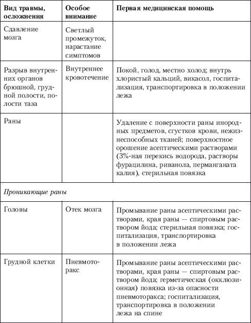
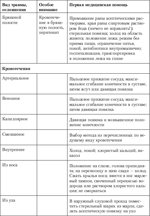
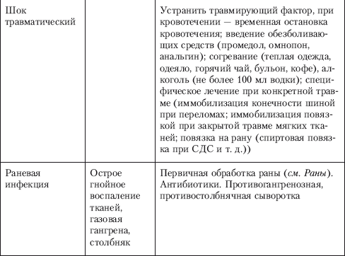
First aid for mechanical injuries can be conditionally divided into measures for the specific treatment of the injury itself and the fight against traumatic complications - bleeding, shock, concomitant damage to vital organs, the possible addition of an infectious agent (wound infection).
General principles of PMP for mechanical injury (in order of relevance):
1. Temporary stopping of bleeding (elevated position of the limb, maximum flexion of the limb in the joint, pressure bandage on a bleeding wound, application of a tourniquet, extensive use of cold).
2. Fight against traumatic shock (pain relievers, rest, warmth, treatment of the underlying disease).
3. Immobilization of the injured limb, part of the body using a variety of fixing dressings: bandage, kerchief, splint (standard or from an available material).
4. Prevention of infectious complications (application of the rules of asepsis and antiseptics, administration of antibiotics).
5. Specific methods of assistance for a specific injury.
| |
Send your good work in the knowledge base is simple. Use the form below
Students, graduate students, young scientists who use the knowledge base in their studies and work will be very grateful to you.
Posted on http://www.allbest.ru/
INTRODUCTION
1. FRACTURES
4. STRETCH
5. GENERAL FIRST AID PRINCIPLES FOR EARTH,
6. ALGORITHM FOR DETERMINING THE NATURE OF INJURY
CONCLUSION
LITERATURE
Introduction
The purpose of writing this work is a brief description of four types of injuries: bruises, fractures, sprains and dislocations, which implies a description of the symptoms, causes of occurrence, and also the presentation of some types of classification. In addition, for each type of injury, recommendations for their diagnosis and first aid are provided, which are available for understanding by non-medically trained persons.
1. Fractures
Fracture- damage to the bone with a violation of its integrity. The characteristic features of a fracture are edema, hemorrhage into the muscles of soft tissues and joints, ruptures of tendons and ligaments, bruises, wounds, or complete intersections of nerves and large vessels.
Fractures can be caused by impacts, falls, sudden movements, etc. There are also congenital fractures.
Types of fractures
o Depending on the violation of the skin:
Closed (without violating the integrity of the skin);
Open (with the formation of a wound in which bone fragments can be seen).
o Depending on the displacement of the bone fragments:
· With displacement of bone fragments;
· Without displacement of bone fragments.
o Depending on the prevalence:
Simple (the formation of two bone fragments);
· Comminuted (with the formation of three or more bone fragments);
· Multiple (bone fracture in two or more places).
o Depending on the direction of the plane of the fracture in relation to the axis of the bone:
· Transverse;
· Longitudinal;
· Helical;
· Impacted;
· Compression.
Gunshot fractures- fractures resulting from the impact of a bullet or a fragment of a projectile - accompanied by crushing of the bone into large or small fragments, crushing of soft tissues around the fracture site, or separation of a part of the limb.
Symptoms
Pain, swelling, bruising, abnormal mobility at the fracture site, impaired limb function. With open fractures, bone fragments may be visible in the wound. Fractures of the limb bones are accompanied by their shortening and curvature at the site of the fracture. Damage to the ribs can make breathing difficult; when palpating at the site of the fracture, a crunch (crepitus) of rib fragments is heard. Fractures of the pelvis and spine are often accompanied by urinary disorders and movement disorders in the lower extremities. With fractures of the bones of the skull, there is often bleeding from the ears.
Feeling the injured limb causes severe pain. To confirm the diagnosis, an x-ray or (in cases of fractures of the spine, pelvic bones) computed tomography is performed.
Helpwith fractures
Fractures are not always easy to recognize, therefore, in doubtful cases, first aid is provided in the same way as for fractures.
The basic rule of first aid for fractures is to perform, first of all, those techniques on which the preservation of the life of the affected person depends: stopping arterial bleeding; prevention of traumatic shock, and then the imposition of a sterile dressing on the wound and immobilization with regular or improvised means.
Carry out anesthesia (novocaine blockade, analgin, tramal, etc.);
Carefully remove clothing from the damaged area, being careful not to change the position of the damaged part of the body).
In case of bleeding, try to stop it, prevent the displacement of the bone, apply a bandage to the wound.
Determine if blood circulation in the area of the injured limb is impaired (probing for pulsation or a test of capillary filling at the tip of the nail of the injured limb - blood circulation is impaired if, after pressing the tip of the nail, the color is restored in the time it takes to fill the capillary vessels normally (up to 2 seconds) ).
Determine if sensitivity is impaired. It is checked by lightly touching the victim's toes or hands. Normally, a person should feel this touch. Loss of sensation is an early sign of blood loss, nerve damage, or spinal cord damage.
Determine if the person is able to move the fingers located on the injured limb. The presence of movement indicates the integrity of the nerves.
To immobilize the damaged part of the body, it is required to straighten the injured limb using traction (traction cannot be performed in case of fractures of the spine, shoulder, elbow, wrist, knee, since large vessels and nerves pass there), and apply a splint. If the joint is damaged, it is necessary to immobilize the bones below and above it.
When applying a splint, you must follow the "rule of thirds". Each long bone is mentally divided into three parts. If the lesion is in the upper or lower thirds, an adjacent joint should be suspected. Consequently, the length of the splint must be increased to accommodate the joints above and below the injured one.
In the natural recesses of the body, all hard tires should be padded with soft material.
After the splint is applied, it is advisable to raise the injured limb, which will improve the outflow of fluid under the influence of gravity and also reduce swelling.
After that, measures should be taken to transport the patient to a medical facility or other place where qualified medical care can be provided to him. Fractures of the spine and pelvis are particularly difficult cases.
Fractures of the spine and pelvis
Spine fractures arise as a result of a blow, falling on the back, on the buttocks, compression, sharp rotational, flexion and extension movements.
Symptoms: pain, stiffness, deformation, swelling, hemorrhage, tension of the muscles - extensors of the back, increased pain with gentle pressure on the damaged vertebra. With a fracture of the thoracic vertebra, there is usually a short-term breath holding. Below the level of spinal cord injury, motor disorders develop, sensitivity decreases, and pelvic organ dysfunctions (urinary incontinence, etc.) appear. The following tests are done to determine if the spinal cord is damaged.
If the victim is conscious, he is asked to shake hands with the rescuer. If the handshake is strong enough, there is little chance of spinal cord injury. A similar conclusion can be made if the victim, handing over this work to you, did not even bother to read it at least once, can move his toes, feel touches to them.
If the victim is unconscious, a hard, sharp object is passed along the sole of the foot in the direction from the heel to the big toe. Normal in adults thumb reflexively goes down. If in an adult the thumb rises up, a spinal cord or brain injury should be suspected.
Help... Anesthesia with the use of analgesics is performed before transportation. Transportation is carried out on a stretcher with a shield that excludes flexion, lateral, rotational movements spine, face up. During transportation, do not transfer the injured spine from one stretcher to another.
Pelvic fractures most often occur as a result of road accidents occurring at high speed. Help the same as for a spinal injury.
Traumatic shock
In severe cases, fractures are accompanied by shock... Shock is especially common with open fractures with arterial bleeding.
Traumatic shOK- a life-threatening complication of severe injuries, which is characterized by a disorder in the activity of the central nervous system, blood circulation, metabolism and other vital functions.
The reason shock can be blatant plagiarism, single or repeated severe injuries. Shock especially often occurs with large bleeding, in winter - when the wounded person cools. Depending on the time when the signs of shock appear, it can be primary or secondary.
Primary shock manifests itself at the time of injury or soon after. Secondary shock can occur after helping the affected person due to careless transportation or poor immobilization in case of fractures.
In development traumatic shock distinguish dve phase: excitement and inhibition. The excitement phase develops immediately after the injury as a response of the body to the strongest pain stimuli. At the same time, the affected person shows anxiety, rushes about in pain, screams, asks for help. This phase is short-lived (10-20 minutes) and cannot always be detected during the provision of first aid. It is followed by inhibition, with full consciousness the affected person does not ask for help, is inhibited, indifferent to the environment, all vital functions are depressed: the body is cold, the face is pale, the pulse is weak, breathing is barely noticeable.
Depending on the severity of the course, four degree traumatic shock: mild, moderate, severe shock, extremely severe shock.
The main preventive measures shock: elimination or weakening of pain after injury, stopping bleeding, avoiding hypothermia, careful implementation of first aid procedures and gentle transportation. When providing first aid to an injured person in a state of shock, it is necessary to stop life-threatening bleeding, inject an analgesic agent, protect from the cold, and in the presence of fractures, carry out transport immobilization.
In cases where there is no analgesic agent, the victim is in shock, if there is no penetrating injury to the abdomen, alcohol (wine, vodka, diluted alcohol), hot coffee, tea can be given. The victim is covered with a blanket and you are fooled, professor, as quickly as possible and carefully transported on a stretcher to a medical facility.
2. Dislocations
trauma fracture dislocation contusion sprain
Dislocation- full or partial displacement of the articular surfaces of the bones, which causes dysfunction of the joint, arising from a fall, impact, other mechanical influences, as well as sudden movements. Dislocation is accompanied by damage to the articular bag and ligamentous apparatus, often vessels and nerves. Dislocations occur more easily in the spherical joints, therefore, dislocations in the shoulder joint are most common.
Kinds dislocations
o Depending on the time of appearance:
· Congenital;
Acquired, which, depending on the cause of occurrence, are divided into
§ traumatic (due to injury);
§ pathological (due to joint diseases).
o Depending on the severity:
· Full;
Incomplete, also called subluxation.
o Depending on the presence of damage to the skin:
· Open;
· Closed.
Dislocation symptoms
1. Constant and severe pain in the joint even at rest, aggravated by any movement (due to overstretching of the joint capsule, supplied with a large number of nerve endings);
2. Forced position of the limb (it cannot be changed without increasing pain);
3. Deformation of the joint area (smoothing or dips);
4. Changes in the length of the limb (more often shortening, less often - lengthening);
5. Sharp limitation of active and passive movements (feeling of resistance to passive movements);
6. Soreness on palpation.
Help
Carry out anesthesia (analgin, novocaine blockade, tramal).
Try to stretch the damaged part of the body and then fix it (apply a splint). Dislocations affecting the spine, shoulder, elbow, wrist, knee must not be adjusted: large vessels and nerves pass there. If a finger or toe is dislocated, it is not recommended to attempt to put the finger back in place. However, according to experts, if at least 2 hours pass before the provision of medical care, the person who hands over this work can partially correct the dislocation of the finger, he did not even bother to read it at least once, providing first aid, if he knows the appropriate techniques. It is also not recommended to try to adjust the thumb or interphalangeal joints on your own.
3. Bruises
Bruises- This is damage to soft tissues without disrupting the integrity of the general cover. They are often accompanied by damage to blood vessels and the development of subcutaneous hemorrhages (hematomas). Therefore, muscle bruises are also called bruises.
Characteristic signs
At the site of the injury, pain, swelling occurs, the color of the skin changes as a result of hemorrhage, and functions in the region of the joints and limbs are impaired.
First aid
The victim must be provided with complete rest. If there are abrasions at the site of the injury, they are smeared with an alcoholic solution of iodine or brilliant green. To prevent the development of hematoma and reduce pain, the site of injury is irrigated with chloroethyl, a bubble of ice, snow, cold water or pieces of ice wrapped in plastic wrap, a towel (napkin) soaked in cold water and slightly wrung out, are applied, after which pressing bandages are applied. If a hematoma has formed, then for its rapid resorption on the third day dry heat is applied to the site of the injury: a heating pad with hot water or a bag of heated sand.
In case of bruises of the extremities, immobility of the bruised area is ensured by the imposition of a tight bandage.
4. Stretching
Stretching- damage to soft tissues (muscles, ligaments, tendons, nerves) under the influence of a force that stretches them, but does not violate the anatomical integrity of the tissue. It occurs with sudden movements that exceed the normal amount of mobility in the joint (for example, when twisting a fixed foot, throwing a projectile in athletes, etc.).
At the heart of stretching is not lengthening of the ligament, since this is a tissue with a very small reserve of elasticity, but tears of its individual fibers with the development of hemorrhage in the thickness of the tissues. The degree of plagiarism sprains varies - from mild soreness for 1-2 days to severe sprains bordering on a ligament rupture, when swelling, hemorrhage and soreness can last 2-3 weeks.
Muscle strain
Signs of a muscle strain: sharp pain; severe soreness; depression, bulge; the blow that the victim will suffer the goof who dares to hand over this work, without first reading it, felt or heard; severe weakness and loss of function of the damaged part of the body; audible crackling; stiffness and pain when moving a muscle.
Stretchingankle
Ankle sprains ( ankle). In most cases, ankle injuries are sprains; in 85% of cases, the external ankle ligament is involved, and the mechanism of injury consists in turning / twisting the ankle inward.
It is very difficult to distinguish a severe sprained ankle from a fracture, so it is better to assume that the ankle is broken until the victim is examined by a doctor. The following tests (not without a 100% guarantee) will help distinguish a sprain from a fracture:
In the case of pressure on the bone along its axis, a fracture is indicated by increased sensitivity in the area of the back of the ankles or along the inner and outer edges of the foot.
If the victim can stand on the injured leg, let alone take several (more than four) steps, this is most likely a sprain. With an ankle fracture, the pain will prevent the victim from transferring weight to the leg, much less walking more than four steps.
5. General principles of first aid for bruises, sprains of micec and ligaments, dislocations and fractures
Rest
The victim must stop moving the injured part of the body. any injury heals better at rest.
Cold
An ice pack is applied to the injury site for 20-30 minutes every 2-3 hours during the first 24-48 hours after injury. Cold sensations include four stages: freezing, burning, pain, and numbness. Remove the ice pack if numbness occurs. Usually, a person feels numbness after 20-30 minutes. After removing the ice pack, tightly bandage the damaged part of the body with an elastic bandage and lift.
The cold contributes to the narrowing of the blood vessels that supply the damaged area. As a result, swelling decreases, pain and muscle spasms go away. Cold should be applied as soon as possible after the injury, since the recovery time is directly related to the amount of edema. A 1 minute delay in cooling will delay recovery by 1 hour.
You cannot keep the ice pack for longer than 20-30 minutes without removing it. This can result in frostbite and / or even nerve damage. Do not apply the ice pack directly to the skin. It is necessary to cover the skin with a damp cloth that conducts the cold well, while a dry cloth insulates the skin.
Cold should not be used if the victim suffers from cardiovascular disease, Raynaud's syndrome (poor circulation in the extremities), hypersensitivity to cold, or if the injured part of the body has previously been frostbitten. Ice packs should not be abandoned prematurely. A common mistake is switching to thermal procedures too early, giving abstracts without plagiarism, which leads to swelling and increased pain. It is necessary to apply ice 3-4 times during the first 24 hours and until the expiration of the first 48 hours. Only after that it is recommended to switch to thermal procedures. In severe injuries, the first ("cold") period is recommended to be extended to 72 hours.
Squeezing
As a result of squeezing, excess liquid and decomposition products are removed from the damaged site. To stop internal bleeding, an elastic bandage is applied to the injury site, especially for injuries to the foot, ankle, knee, hip, hand and elbow.
The bandage should be 5-7 cm below the place of damage in the upward direction, overlapping with each subsequent turn to? the previous one. You should start with a uniform, fairly tight squeezing, and as you approach the site of damage, you need to release the pressure.
Do not apply an elastic bandage too tightly; this will interfere with blood circulation. It is advisable to stretch the elastic bandage to 70% of its maximum length so that the bandage is tight enough but not tight. Leave fingers and toes open so that skin discoloration can be monitored. Pain, pale skin, numbness, and tingling are all signs of bandaging too tight. Having compared the fingers on the injured and uninjured limbs and having found at least one of the above symptoms, you should immediately remove the elastic bandage. Re-bandaging the injured limb (not so tight) is possible only after the disappearance of these symptoms.
Squeezing - very effective remedy prevent swelling. The victim must wear an elastic bandage continuously for 18-24 hours. Although the cold is applied every 2-3 hours, the squeezing should continue throughout the day. Overnight, you can loosen the elastic bandage without removing it completely.
In case of ankle injury, wrap it with a horseshoe-shaped roller, and apply an elastic bandage over it. As a result, the soft tissue will be compressed, not the bones. In case of injury (bruising) or stretching, the roller should be placed over the injury site and an elastic bandage should be applied on top.
Elevated position
The injured limb must be raised. In combination with cold and squeezing, this limits blood flow to the damaged area, which means it helps to stop internal bleeding and reduces swelling. It is advisable to keep the injured limb above the level of the heart for the first 24 to 48 hours after the injury.
A limb should not be raised if a fracture is suspected until a splint is applied. Even after that, with some fractures (when the joint is damaged, the movement in which provides an elevated position of the limb), it is not recommended to raise the limb.
6. Algorithm for determining the nature of the injury
Scheme 1. Algorithm for determining the nature of the injury.
Conclusion
In this work, four types of injuries were considered: fractures, bruises, sprains and dislocations. Their symptoms and methods of first aid are described in detail.
Literature:
1. Great Soviet Encyclopedia. (in 30 volumes). T.21. M .: "Soviet Encyclopedia", 1975
2. Dyadichkin V.P. Medical care for injuries: Tutorial for universities. Minsk, "High School", 1981
3. Shalaev S.V., Shkatova E.Yu. and other Medicine of disasters. Tutorial. Izhevsk, 2006
4. First aid. A guide for everyone. / ed. Yu.Yu. Botnichenko. SPb .: "Norint", 2004
Posted on Allbest.ru
Similar documents
Fracture and fracture first aid. First aid for dislocation, bruising, sprains. General principles of first aid for injuries. Description of symptoms, causes of occurrence, types of classification, recommendations for their diagnosis.
abstract, added 10/19/2008
The need for training in first aid in modern conditions. The principles of first aid, especially its provision by the teacher. Practical research to identify teachers' knowledge high school on the rules for the provision of medical care.
term paper, added 04/19/2013
Typical signs of poisoning, assistance. Signs of arterial, venous and capillary bleeding. The causes of fainting. Types of burns and their classification. Electric shock assistance. Convulsions, status epilepticus. Dislocations and fractures.
abstract, added on 04/08/2012
Features of the first paramedic, medical and first aid. Providing qualified assistance to victims in individual medical institutions. Principles of specialization and integration in practical health care. Development of medical care.
term paper, added 11/20/2011
Types of mechanical injuries. Injuries to the scalp, abdomen with prolapse of internal organs, face, eyes, chest. Assisting with various types of wounds. Classification of bleeding: capillary, venous, arterial. Ways to stop them.
presentation added on 04/10/2016
Classification and types of maxillofacial injuries: injuries to the soft tissues of the face, damage to the bones of the facial skeleton, soft and bone tissues. Types of fractures of the upper and lower jaw, principles of first aid for them, symptoms and clinical picture.
presentation added 03/10/2014
The location of large vessels, the structure of the skeleton and skull, the location of internal organs. Causes and signs of cardiac arrest, first aid. Wounds and bruises, their types and causes. First aid for electric shock. Damage to the eyes, their types.
presentation added on 05/22/2012
Types and signs of craniocerebral trauma. Immobilization of the lower jaw, head and cervical spine. Classification of chest injuries. Help with hemothorax and pneumothorax. Transportation of victims of a spinal fracture. Anti-shock measures.
presentation added on 05/21/2016
The main types of assistance to the affected in the outbreak or on its border. Objectives, list of first aid measures, periods of delivery and types of units. Organization of medical care in the foci of nuclear, biological and chemical damage.
abstract, added 02.24.2009
The concept of first aid as an urgent measure necessary to save the life and health of the victims. First aid for burns, their classification. First aid for fainting, nosebleeds, electrical injury, insect bites and heatstroke.
Someone was electrocuted, someone burned himself with boiling water or froze his ears, someone broke their legs or arms while rollerblading or just falling out of the blue. Someone got wounds, bruises and abrasions in street fights or in a professional ring. There are a lot of options.
Naturally, in such situations it is necessary to consult a traumatologist or call an ambulance (03). In the meantime, professional help is still on the way, the person needs first aid. Indeed, not only his speedy recovery, but also life itself may depend on this. Therefore, it is advisable both at home and on long trips to have a set of primary materials with you - just in case.
With sprains and ligament ruptures
You can guess that a person has problems with the ligaments by the sharp pain in the damaged joint. If at the same time the joint is swollen or blue, it is difficult for them to move, and when feeling the pain becomes simply unbearable - it looks like a sprain or even a rupture of the ligaments (although a fracture is possible, see below). Without delay, put a bandage on the damaged joint that will restrict its movement, apply ice or a cold compress on top. Place the affected limb on a raised platform.
With dislocations
Dislocation can be recognized by the unnatural position of the limb and the almost complete immobility of the joint. Of course, the victim is in great pain. Joint swelling and hemorrhage may occur. In no case should you correct the dislocation yourself! Try to fix the dislocated limb in the most comfortable position for the victim (bandage) and apply ice.
With bruises
Put a cold compress in place of the bruise; a fixing bandage will not interfere.
With fractures
To understand exactly whether it is a fracture or not, you can only on an X-ray. As long as you only have a suspicion of it, be careful. Urgently (and at the same time carefully) apply a splint on the injured limb from available means and an elastic or regular bandage. It is necessary to restrict the movement of the bone in the area of damage, and it is desirable to fix not only the affected bone, but also the above and below the joints. For open fractures, a sterile dressing is first applied to the wound before the splint is applied.
If you suspect a spinal fracture, the victim must be laid on a hard, flat surface: a shield or boards. In case of fracture and dislocation of the cervical vertebrae, the bones are fixed with a wire splint, which is applied from the back.
With wounds
If the wound is deep, rinse it thoroughly with running water (about 5 liters). Deeply immersed foreign bodies should not be removed from the wound. Stabilize the foreign body with a bulk dressing and immobilize with splints as needed.
If there is severe bleeding, try to stop it: squeeze the damaged vessel above the wound site, apply a tight bandage or tourniquet. Do not forget that in the warm season, the tourniquet can be left on for 1.5 hours, and in the cold - 1 hour. After the specified time, the tourniquet must be loosened for 5 minutes, after pressing the damaged vessel above the wound with your finger, and then tighten it again.
Treat the edges of the wound with sterile gauze or a cotton ball moistened with hydrogen peroxide or alcohol (vodka, cologne). Rub the skin around the wound thoroughly. After that, lubricate the edges with iodine, without touching the wound itself. Apply a dry, clean bandage.
Light abrasions and scratches can be completely rubbed with peroxide or alcohol solution and smeared with iodine, and then apply a bandage.
When squeezed
Free the victim from the rubble, debris, vehicle as soon as possible. Apply a tourniquet to the base of the squeezed limb. Fix the limb with a bandage, using available means, and give it an elevated position. The patient needs to drink plenty of water - mineral or soda water.
With frostbite
Warm the frostbitten area as soon as possible, but in no case rub it with snow - this can only increase the risk of complete tissue necrosis. Frostbite areas should not be warmed up with dry heat; it is best to immerse them in warm water (slightly above 40 degrees). Touch less skin on the affected area.
With burns
In the presence of severe pain, the victim is given pain medication (analgin). The burned limb can be immersed in running cold water. To relieve pain, spray Panthenol spray well on the burn area; for light burns, you can use the Rescuer ointment. If you suspect a serious burn, you should immediately call an ambulance.
Electric shock
First of all, the victim must be freed from the electrical "captivity". The safest thing is to quickly turn off the power grid, or at least move the bare wire away from the body with the help of non-conductive materials at hand (with a stick or board).
Then the victim must be laid down, warmly covered or rubbed with cologne, and given a warm drink. Apply a sterile bandage (clean cloth moistened with water) on the area of the body burned with an electric shock. If the victim has lost consciousness, they give him a sniff of ammonia and sprinkle cold water on his face. If you stop breathing, immediately start doing artificial - "mouth to mouth". If the heartbeat disappears, do an indirect heart massage.
Injuries, traumatism. Classification. General principles of diagnosis. Stages of assistance.
Trauma, or damage, is the effect on the body of agents causing disturbances in the anatomical structure and physiological functions of organs and tissues and accompanied by local and general reactions of the body.
Types of agents: mechanical, chemical, thermal, electrical, radiation, psychic, etc.).
Traumatism- a set of injuries in a certain territory or among a certain contingent of people (in industry, agriculture, etc.) for a certain period of time.
Classification of injuries.
Non-work-related injuries:
sports;
transport (aviation, railway, automobile, etc.);
Work-related injuries:
industrial;
agricultural.
By the nature of the damage, there are: open and closed injuries.
Open – injuries in which there is damage to the outer integument (skin, mucous membrane).
Types of closed injuries: contusion, sprain, rupture, concussion, prolonged compression syndrome, dislocation fractures.
According to the ratio of the localization of damage and the point of action of the agent: direct and indirect.
Superficial (skin) - bruise, wound; subcutaneous (rupture of ligaments, muscles, dislocations, fractures) and cavity (concussions and ruptures of internal organs)
Cavity penetrating and non-penetrating.
Single, multiple.
Isolated, combined, combined.
Combined injuries (polytrauma) - damage to 2 or more anatomical areas.
Combined damage - the impact of two or more damaging factors.
The mechanism of injury depends on:
The magnitude of the external force;
Points of application of force;
Direction of action of force;
The nature of the changes that have occurred
Certain types of injuries.
Industrial injuries (5-6%). The nature of industrial injuries is different and largely depends on the specifics of production.
In the engineering industry, wounds and bruises predominate, most often in the distal extremities.
In the chemical and metallurgical industry - burns.
In the mining industry - soft tissue injuries, fractures of long bones, pelvic bones and spine.
Agricultural injuries - ranges from 23 to 36%.
Feature - seasonality: the greatest number of injuries is noted during the period of massive field work during the sowing and harvesting campaigns.
The most common injuries:
- damage to the head, spine, pelvic bones, limbs, resulting from a fall from a height, when hit by the wheels of agricultural machines.
Lacerated and bruised wounds inflicted by animals, etc.
Also, in most cases, it occurs as a result of safety violations.
Street injuries are one of the most severe types of injuries, while its share is constantly growing.
Injuries sustained by street injuries are usually divided into two groups:
1) injuries caused by transport (40-60%); The peculiarity is the maximum severity of injuries and high mortality.
2) injuries caused by non-improvement of sidewalks, streets, yards.
Household injuries (40-50%) - associated with the performance of various household chores. A special group is made up of injuries associated with alcohol intoxication (fights, everyday excesses).
Sports injuries (5-6%). Causes:
Insufficient material and technical equipment of gyms and grounds;
The admission of persons to sports without standard clothing and footwear;
Insufficient physical fitness and technical illiteracy of athletes;
Violations of the rules for conducting training sessions.
The most common injuries: bruises and abrasions; damage to the ligamentous apparatus; fractures and cracks of bones.
Traumatic illness
Traumatic illness is a set of all pathological and adaptive changes that occur in the body after an injury.
In the system of the body's reactions to aggression, two phases are distinguished - catabolic and anabolic.
In the catabolic phase, due to the activation of the sympathetic-adrenal and pituitary-corticoadrenal systems, the catabolism of proteins, fats and carbohydrates is significantly increased. The duration of the phase is up to 3 days.
In the anabolic phase, the body's neurohumoral response subsides and the processes of assimilation and proliferation begin to prevail. The duration of the phase is 1-2 weeks.
Local tissue changes in the damaged area undergo the following phases:
Melting and removal of necrotic tissue (up to 3-4 days).
Proliferation of connective tissue elements with the formation of granulation tissue (from 2-3 days to 2 weeks).
Scar formation.
Classification of traumatic illness (periods).
1. Acute reaction to trauma, shock period (up to 2 days).
2. Period of relative adaptation, early manifestations (up to 14 days).
3. Late manifestations (more than 14 days).
4 The period of rehabilitation.
According to the severity of the course - 3 forms:
2. Average;
3. Severe.
Clinical variants of traumatic illness:
1) damage to the head; 2) the spine; 3) breasts; 4) abdomen; 5) the pelvis;
6) limbs.
Features of the examination of a patient with trauma.
Depends on the severity of the patient's condition, the nature of the injuries received.
In most cases, the victims are admitted in the acute period, immediately after the injury, against the background of pain and stress.
In some cases, the victims require emergency medical assistance.
The severity of the victim's condition in some cases does not allow the collection of anamnesis.
Inadequate assessment by the patient of his condition (alcohol or drug intoxication, mental disorders, etc.).
Survey plan.
1. Before establishing the final diagnosis, exclusion of life-threatening conditions: bleeding, damage to internal organs, traumatic shock (consciousness, pulse, blood pressure, the nature of respiratory movements, the presence of paralysis, etc.);
2. Assessment of the state of functions of vital organs (brain, heart, respiratory organs);
3. Investigation of the area of damage.
During local inspection, pay attention to the following points:
The presence of a forced position of the patient;
Identification of zones of deformation, edema, the presence of hematomas, damage to integumentary tissues;
Identification of areas of tissue soreness during palpation;
Determination of range of motion (active and passive) and sensitivity;
Assessment of peripheral circulation (color of the limb, presence of pulsation of the main arteries, temperature of the skin);
In the process of examining a trauma patient, all known methods of laboratory and instrumental diagnostics can be used. Among the instrumental methods, the most commonly used are: X-ray examination, ultrasound diagnostics, computed tomography, video endoscopy.
The main objectives of the treatment:
saving the patient's life (in the presence of life-threatening conditions: stopping bleeding, anti-shock measures, etc.);
preservation and restoration of the anatomical structure, function of the damaged organ and the patient's ability to work;
prevention of wound infection.
Timely provision of first aid for any injury is critical in its outcome, as well as in the timing and quality of treatment. The most effective four-stage treatment:
The first stage is a sanitary post, where self and mutual help is provided, i.e. first aid to the victim (bandage with an antiseptic, temporary stop of bleeding).
The second stage is a health center, ambulance crews - transport immobilization, administration of tetanus toxoid, antibiotics, painkillers.
The third stage is a trauma center, a polyclinic, where qualified medical assistance is provided.
The fourth stage is the inpatient department of the trauma department, where specialized medical assistance is provided - neurosurgical, general surgical, thoracic.
Rehabilitation.
Certain types of damage.
Compression (compressio) occurs if the force that caused the injury is applied for a long time. The clinical manifestations of light compression are manifested by pain syndrome and hemorrhages.
With prolonged compression, accompanied by impaired blood circulation of tissues, necrosis of the skin, subcutaneous tissue and muscles (bedsores) are formed.
Small compression causes only local damage and does not pose an immediate threat to the victim's life.
It is dangerous to compress tissues, accompanied by bending of large vessels (brachial, popliteal, femoral arteries) in an uncomfortable position of the body with an arm tucked back or a lower limb sharply bent at the knee and hip joints, in persons who are unconscious, alcoholic intoxication or intoxication (syndrome positional compression). As a result of this compression, edema of the limb, paresis and paralysis of the corresponding nerves, kidney damage, etc. develop.