Radiculitis, or, in other words, radicular syndrome, is one of the manifestations of osteochondrosis: degenerative changes occur in the intervertebral discs, due to which the annulus fibrosus breaks and a hernia forms. It compresses one or more roots of the spinal cord, or it exerts compression on the ligamentous apparatus of the spine. As a result of pinching the roots, radiculitis occurs.
Symptoms of sciatica
In most cases, lumbosacral and cervicobrachial sciatica occurs. The main signs that sciatica has is pain in the lower back, which can radiate to the back of the leg, buttocks, knees or lower leg. If you try to bend forward or sit down with your legs straight, the pain will be much worse. To minimise pain, the patient slightly bends the leg. Along with pain, there is tingling or numbness in the lower leg and fingers. In addition to the pain syndrome, there is a change in the patient's posture, curvature of the spine.

Radiculitis, regardless of the location, have similar symptoms: the appearance of rapid pain in the area of the affected roots, which increases when the patient moves, coughs or sneezes, stiffness in the mobility of the spine; pain on palpation of the spinous processes of the vertebrae and paravertebral points; increase or decrease in sensitivity; muscle weakening in the area of radicular innervation.
The pains that accompany sciatica are usually shooting, breaking, increasing when raising the leg, coughing, hypothermia. Sciatica can recur, accompanied by tension on the nerves and roots, the presence of pain points and impaired sensitivity. Lumbosacral sciatica is characterized by the appearance of pain throughout the day, regardless of the time, increase with a change in the position of the body.
Radiculitis treatment
If you have sciatica, bed rest must be strictly observed. Analgesics are used to reduce pain. Before getting out of bed, you need to fix the patient's lower back with a special belt, in the lying position it should be removed.
Blocks of novocaine, lidocaine and vitamin B12 in pain points have a positive effect. At night, you can apply a compress of Dimexide diluted with water, novocaine, analginum, vitamin B12 and hydrocortisone on the lumbar region.
Indomethacin is taken internally. To eliminate muscle tension that accompany sciatica, it is advisable to take seduxen, diazepam. Also shown is a relaxing massage of the back and buttocks. The massage should be performed by a professional so as not to injure the patient with careless movements. Sciatica can also be relieved with acupuncture and physiotherapy using current, ultrasound, etc.
Radiculitis can be calmed with the help of heat on the lumbar region (heating pad, paraffin application), mud therapy is practiced, the use of salt-pine baths. For prevention, hardening of the body, limiting physical activity, hypothermia, and long walking are also recommended.
Traction treatment, or traction of the spine, positively affects the receptors of the damaged ligaments of the spine and muscles, relaxing them. This method is widely used during the rehabilitation period after you have practically cured sciatica and has the following effect: unloads the spine, increasing the space between the segments spinal column; reduces muscle tension; lowers pressure inside the disc, and also relieves compression on the nerve roots.
Prophylaxis
In order to prevent sciatica, it is recommended to perform exercises that strengthen the muscles of the back, swim, avoid hypothermia, physical overload. The main task physical exercise in the treatment of sciatica - to help normalize muscle tone back, increase the mobility of the spine, improve overall well-being and accelerate the process of rehabilitation and recovery labor activity... The set of exercises is selected based on the symptoms of the disease, the general condition and age characteristics of the patient.
Radiculitis is a fairly common disease of the peripheral nervous system, which is formed as a result of compression of the roots of the spinal cord. Only a specialist can prescribe treatment and conduct an examination. For the most accurate determination of the diagnosis of sciatica, the doctor will first determine muscle strength, differentiate the symptoms, the nature of pain, their intensity, duration, determine if there is a sensitivity disorder, prescribe X-ray or other examination methods, after which complex treatment will be prescribed.
Medical Expert Editor
Alexey Portnov
Education: Kiev National Medical University. A.A. Bogomolets, specialty - "General Medicine"
radiculitis
Radiculitis is a disease of the roots of the spinal cord. Distinguish between primary (infectious, toxic) and secondary radiculitis, most often associated with pathology of the spine and spinal cord. There are also meningoradiculitis - infection roots with the spread of the pathological process to the membranes of the spinal cord; occurs with rheumatism, brucellosis, syphilis and is manifested by multiple lesions of the roots and meningeal symptoms.
Secondary radiculitis can be caused by root neuromas, spinal tumors, tuberculous spondylitis, osteomyelitis, osteochondrosis, traumatic spinal injury. Radicular pain in neuromas is permanent, does not respond to drug therapy and is combined with signs of spinal cord compression and an increase in the protein content in the cerebrospinal fluid (see Spinal cord, tumors). In women, sciatica is often caused by inflammation or swelling of the ovaries and uterus.
The most common cause of secondary radiculitis is spinal osteochondrosis (spondylosis, spondyloarthrosis), i.e. age-related degenerative changes in the intervertebral discs, joints and ligaments of the spinal column. The process begins with dehydration of the discs, the nucleus pulposus of which loses its elasticity and through the cracks in the fibrous ring can bulge outward, which is accompanied by the formation of a herniated disc (see Lumbago). As the discs degenerate, bone spines - osteophytes - grow at the edges of the vertebral bodies. Herniated discs and osteophytes most often occur in the lumbar and cervical spine, mainly between IV and V lumbar, V lumbar and I sacral and between V-VI and VII cervical vertebrae... Discogenic radiculitis develops in people engaged in heavy physical labor, or in untrained people with congenital weakness of the ligamentous-articular apparatus of the spine.
Clinical picture primary and secondary radiculitis has a number of similarities, but secondary radiculitis are more common.
Acute and chronic radiculitis are distinguished along the course, the latter often being of a recurrent nature. Sciatica is manifested by local pain along one or more closely located posterior nerve roots. Along with pain, sensory disturbances are observed, less often movement disorders. During lumbosacral sciatica there are two stages: lumbar and radicular.
Lumbodynia is a dull aching or sharp pain in the lumbar region that occurs after prolonged physical work, especially in an uncomfortable position of the body and in the cold, or with awkward movement. The lumbar stage is associated with reflex or mechanical irritation of the nerve endings in the ligamentous apparatus of the spine. To reduce pain, the patient takes a forced fixed position with the torso tilted forward or to the side, avoiding even the slightest movements of the spine, especially in the lumbar region. When examining a patient in the lower thoracic and lumbar region, a sharp tension of the long muscles of the back is determined, the palpation of which is painful, especially on the diseased side. The duration of this stage ranges from several days to several weeks, after which the pain subsides.
If the disease progresses and passes into the radicular stage, pain spreads from the lumbar region to the buttock, posterior-outer surface of the thigh and lower leg, and can "give" to the heel or thumb. The pains are both one-sided and two-sided. Discogenic radicular pain often occurs after cooling, colds, which cause swelling of the root and pinching it in the disc herniation or osteophyte of the intervertebral foramen. Radicular pains are dull and sharp, accompanied by a burning sensation, "creeping creeps", "passing electric current»In the area innervated by the affected root. The pain increases when walking, sitting, in an upright position, and decreases when lying down. In bed, the patient usually takes a forced position on the side or on the back with the leg bent and brought to the stomach or the knee-elbow position on the stomach, since in these positions the intervertebral openings expand, the tension of the roots decreases and the pain weakens. Sitting on a chair, the patient leans only on the healthy side, and when walking, he drags the injured leg, avoiding sudden movements.
On examination lumbar of the spine during the radicular stage of radiculitis, the lateral curvature of the spine is determined. The calf muscles of the diseased leg lose their tone and become soft, later the muscle loss of the lower leg and thigh develops, paralysis of the extensor muscles of the foot and fingers may appear, the Achilles reflex decreases or is absent. In the course of the affected roots, a decrease in sensitivity is determined. The tension of the nerve trunks causes pain tonic reflexes. These include: Lasegue's symptom, i.e. the appearance of pain when raising a straightened leg up in the patient's supine position, while flexion of the leg in knee joint leads to a decrease in pain; Bonne's symptom - pain when bringing the leg bent at the knee and hip joint; ankylosing spondylitis - pain on the side of the lesion when lifting up a healthy leg in a supine position (cross Lasegue symptom); Neri's symptom - pain along the affected root when bending the head in the position of the patient on his back. The spinous processes of the IV and V lumbar vertebrae (back points of the Gar) are painful when pressed, and when palpating, pain occurs along the midline of the abdomen below the navel (anterior points of the Gar).
Thoracic sciatica manifested by pain along the intercostal roots. With a viral lesion of the intervertebral nodes, "shingles" develops - sharp intercostal pains, aggravated by inhalation, and blistering rashes on the skin in the projection of the affected node.
Cervicobrachial sciatica accompanied by pain along the posterolateral surface of the neck with spread to the spine, scapula, arm, axillary region. The pain increases with bending and turning the head, raising the arm above the horizontal level and abducting it behind the back. When palpating, the paravertebral points in the cervical spine, the nadi subclavian cavities, the inner surface of the shoulder and forearm along the neurovascular bundle are painful. There is a feeling of numbness, burning or tingling in the arm and shoulder girdle along the affected roots, sensitivity decreases, and reflexes with the biceps and triceps muscles decrease and atrophy develops in the muscles of the hand. The hand becomes edematous, cyanotic, cold, pulsation on the radial artery may decrease. Neurogenic tissue damage often develops shoulder joint, which is manifested by swelling, pain and limitation of movement in the shoulder with the development of contracture in the adductor muscles of the shoulder.
Radiculitis treatment: inside amidopyrine (pyramidone), analgin 0.25 g 4 times a day, butadion 0.15 g 3 times a day, with infectious radiculitis penicillin intramuscularly 200,000 IU 4 times a day; local application of dry heat in the form of heating pads, bags of heated sand, ironing through a flannel with a warm iron; rubbing with alcohol, burning ointments (snake venom, bee venom, alcohol with chloroform). For discogenic radiculitis, it is recommended to rest in bed on a flat and hard mattress with a round roller under the lower back; traction treatment, in which the patient is fixed with straps to the raised head end of the bed by the shoulders and chest. The traction by its own weight is carried out for 30-40 minutes. 3-4 times a day. With sharp pains, subcutaneous and intramuscular injection 0.25-2% solution of novocaine, 2-3 ml in the places of greatest pain, intramuscular administration of vitamins: thiamine chloride (B1) - 5% solution of 1 ml and cyanocobalamin (B12) 200 mcg daily. Shown are massage and remedial gymnastics (flexion, extension, lateral torso tilts - with lumbosacral radiculitis, shoulder abduction, head turns - with cervicothoracic radiculitis). Movements should be smooth, gradually increase in volume, and not leave behind painful sensations. Treatment with quartz, ionogalvanization is recommended. In case of chronic recurrent radiculitis, it is necessary to have a spa treatment at mud and balneological resorts. Persistent and often recurrent radiculitis is treated promptly.
Radiculitis (from Latin radicula - root) is an inflammation of the roots of the spinal cord.
The defeat of the roots can occur at different segments along their course from the spinal cord to the exit from the spinal canal. The anterior and posterior roots, upon exiting the spinal cord, pass the subarachnoid space and converge on its lateral surface, where they go further through special openings in the hard and arachnoid membranes, surrounded by sheaths, which are a kind of diverticula of the subarachnoid space; both roots are separated from each other in this place. This part of the roots J. Nageotte called the radicular nerve. The subarachnoid sheath, deeper around the posterior root than around the anterior one, reaches the sensitive ganglion in the lumbar segments. In their further course, the roots are separated from the subarachnoid space and radicular sheaths by fibrous tissue of the dura mater. This part of the roots Nagotte calls the mixed radicular nerve. Sikard (J. Sicard) called this part of the nerve and its continuation to the plexus cord (funiculus). The mixed radicular nerve is placed in the epidural space and the intervertebral foramen (Fig. 1).
The defeat of the roots, surrounded by soft membranes and washed by the cerebrospinal fluid, begins with the primary defeat of the membranes. Therefore, the defeat of this part of the roots is called meningoradiculitis. The defeat of the extrashell part of the roots is called radiculitis. Sicard proposed the term "funicular", which is identical in content. Both of these terms are used in Soviet medical literature.
The indicated localizations of the pathological process differ mainly in the etiology and frequency of damage to one and the other segment of the roots, to a lesser extent in their symptoms.
Neuritis and sciatica:. shoulder NOS. lumbar NOS. lumbosacral NOS. chest NOS Radiculitis NOS Excludes: neuralgia and neuritis NOS (M79.2) radiculopathy with:. lesion of the intervertebral disc cervical(M50.1). lumbar and other intervertebral disc involvement (M51.1) spondylosis (M47.2)
Russian Ministry of Health protocols for diagnosis and treatment
Complex of diagnostic and treatment measures for M54.1 Radiculopathy
Active ingredients for treatment M54.1 Radiculopathy
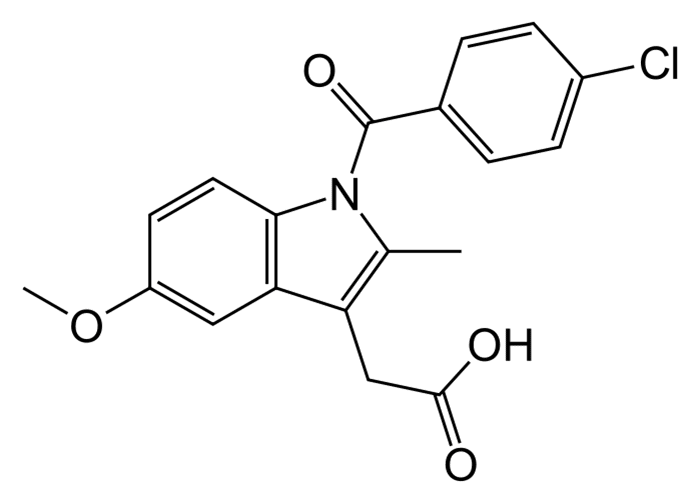
C 21 H 18 ClNO 6
Acemethacin, an ester of glycolic acid of indomethacin, acts as a prodrug. In the body, it is metabolized to indomethacin.
By blocking COX, it disrupts the synthesis of prostaglandins and the production of ATP. It has anti-inflammatory, antipyretic and analgesic effects, inhibits platelet aggregation.
The analgesic effect is due to both central and peripheral effects.
Affects ...

C 19 H 16 ClNO 4
NSAID, a derivative of indoleacetic acid. It has anti-inflammatory, analgesic and antipyretic effects. The mechanism of action is associated with inhibition of the COX enzyme, which leads to inhibition of the synthesis of prostaglandins from arachidonic acid.
When administered orally and parenterally, it helps to relieve pain, especially joint pain in ...

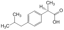
NSAIDs, a derivative of phenylpropionic acid. It has anti-inflammatory, analgesic and antipyretic effects.
The mechanism of action is associated with inhibition of the activity of COX, the main enzyme of the metabolism of arachidonic acid, which is a precursor of prostaglandins, which play a major role in the pathogenesis of inflammation, pain and fever. The analgesic effect is due to both peripheral (indirectly, ...

NSAID, a derivative of propionic acid. It has analgesic, anti-inflammatory and antipyretic effects. The mechanism of action is associated with inhibition of the activity of COX, the main enzyme of the metabolism of arachidonic acid, which is a precursor of prostaglandins, which play a major role in the pathogenesis of inflammation, pain and fever.
The pronounced analgesic effect of ketoprofen is due to two ...

NSAIDs, a derivative of naphthylpropionic acid. It has anti-inflammatory, analgesic and antipyretic effects.
The mechanism of action is associated with inhibition of the COX enzyme, which leads to inhibition of the synthesis of prostaglandins from arachidonic acid.
Suppresses platelet aggregation.
Reduces pain syndrome, incl. joint pain at rest and during movement, morning stiffness ...

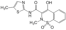
C 14 H 13 N 3 O 4 S 2
NSAIDs, selective COX-2 inhibitor. Belongs to the class of oxicams, is a derivative of enolic acid. It has anti-inflammatory, analgesic and antipyretic effects. The mechanism of action is associated with a decrease in the biosynthesis of prostaglandins as a result of inhibition of the enzymatic activity of COX. At the same time, meloxicam more actively affects COX-2, which is involved in ...

Reversible anticholinesterase agent. Facilitates cholinergic transmission, enhancing and prolonging the action of endogenous acetylcholine. Provides neuromuscular transmission in skeletal muscles, antagonizes curariform remedies non-depolarizing action. Increases smooth muscle tone internal organs, enhances the secretion of exocrine glands. Causes miosis, reduces intraocular ...

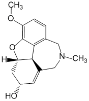
Cholinesterase inhibitor. It prevents the enzymatic hydrolysis of acetylcholine and lengthens its action. Blocks potassium channels of membranes and promotes their depolarization. Stimulates synaptic transmission in neuromuscular endings, conduction of excitation in nerve fibers, enhances the effect on smooth muscles of acetylcholine and other mediators (including epinephrine, ...
A local irritant, which is obtained from a freshwater sponge badyaga, is a colony of coelenterates - Spongilla lacustris fragilis, Ephydatia fliviatilis.
The sponge lives only in exceptionally clean freshwater bodies of water with running water and at certain temperatures.
The badyaga skeleton consists of a looped network of silica needles connected ...
Herbal remedy. Causes moderate sedative, antianginal, carminative, antihypoxic, choleretic, antiseptic, analgesic, antiemetic effects. The healing effects are mainly due to the components of the essential oil, of which menthol is the most studied (60%).
When taken orally (under the tongue), menthol irritates the cold receptors of the oral mucosa, ...
Herbal remedy. Water and alcohol extracts from eucalyptus leaves exhibit bactericidal, antiviral, fungicidal, antiprotozoal and anti-inflammatory effects. Their severity depends on the essential oil content (0.3-4.5%).
The activity of the main component of cineole essential oil (65-85%) is potentiated by pinene, myrtenol, tannins (up to 6%). At...
Eucalyptus oil is obtained from the leaves of various types of eucalyptus. It contains with its composition essential oil, flavonoids, organic acids, tannins and bitter substances, wax, resins. Healing effect means is determined by the totality of the action of the constituent substances.
Eucalyptus oil has a pronounced antibacterial, antiseptic, antiviral effect. It is known that...
Herbal remedy. It has a local warming, irritating and distracting effect, which is provided by capsaicin - an alkaloid-like amide of paprika. The action is aimed at the endings of the peripheral nerves in the skin.
Radiculitis is the most common disease of the peripheral nervous system, in which the bundles of nerve fibers extending from the spinal cord, the so-called roots of the spinal cord, are affected. The most common cause of sciatica is a disease of the spine (osteochondrosis), in which the intervertebral cartilaginous discs lose their elasticity and compress the roots. Salts are deposited at the junction of the vertebrae with the altered discs. The resulting protrusions during exercise put pressure on the nerve roots and cause pain. Sudden movements (turning the torso, head), spasms of the muscles of the back during injury, hypothermia of the body, intoxication can cause the same phenomenon.
The most characteristic manifestations of radiculitis are pain along the affected nerve roots and the nerves formed from them, impaired sensitivity, and sometimes movement disorders. Usually the disease develops acutely, but in many cases becomes chronic with periodic exacerbations. Depending on the location of the lesion of the nerve fibers, they secrete various forms radiculitis. The most common lumbosacral sciatica, in which pains are localized in the lumbosacral region, the buttock with a return to the thigh, lower leg, foot. The pain increases with movement, so the patient avoids sudden movements. In bed, the patient usually flexes the leg to relieve pain.
Lumbosacral radiculitis with a predominance of lesions of the roots of the sacral region, from which the sciatic nerve is formed, is called sciatica. With sciatica, the pain spreads along the course sciatic nerve(in the buttock, posterior outer surface of the thigh and lower leg, heel); accompanied by sensations of coldness of the leg, numbness of the skin, "creeping".
With cervicobrachial sciatica, the pain is noted in the back of the head, shoulder, scapula, and increases with turning the head, moving the arm, and coughing. In severe cases, there is numbness, burning and tingling in the skin of the hand; sensitivity is disturbed. Thoracic sciatica is quite rare and is manifested by pain in the intercostal spaces, aggravated by movement. Treatment is carried out by a doctor; it is aimed mainly at eliminating the causes of sciatica. Physiotherapeutic procedures are widely used along with painkillers, remedial gymnastics, traction of the spine. The independent use of thermal procedures and painkillers is unacceptable, since lower back pain can be caused not only by sciatica, but also by other diseases in which the use of heat is contraindicated. When using funds traditional medicine this must be taken into account. The disease is difficult to cure, and most often a person has to adapt to it. For exacerbations of sciatica, bed rest is recommended.
, 237 ml
The code: A807Colloidal phyto-formula Argo to restore and maintain joint function. Argo company. The main task of the colloidal formula "Arthro Complex" is to help the body with joint diseases: to alleviate the condition (relieve pain, swelling of the joint, restore painless movement, eliminate the "crunch"), increase endurance in articular pathology, reduce the dosage of anti-inflammatory drugs (relieving pain, these drugs stimulate the destruction of cartilage), and in some cases ...
, 100 caps.
The code: RU1295Since time immemorial, Boswellia has been highly regarded for its anti-inflammatory properties. In India, boswelia is called a "fighter against inflammation." Experiencing significant loads (lifting weights, sports), the human musculoskeletal system constantly needs nutritional support. Various diseases of the musculoskeletal system occur against the background of inflammatory processes in the joints. Boswellia Plus has a pronounced anti-inflammatory effect, helps to strengthen and restore st ...
, 60 tablets
The code: V03771Bad Vita B-Plus from Vitaline is a complex of B vitamins. It is necessary for the normal functioning of the central and peripheral nervous system, affects the conduction of nervous excitement in synapses, and due to the presence of vitamin B12 is a growth factor, stimulates erythropoiesis, participates in synthesis of hemoglobin. ...
, 25 g
The code: A1201Recovery and stimulation of metabolic and regenerative processes; prevention and treatment of acne, fungal infections and herpes; healing of trophic ulcers, erysipelas, boils; elimination of puffiness, allergic and other irritations. ...
, 25 g
The code: A1209For massage with acute and subacute symptoms of osteochondrosis and radiculitis, as well as with neuralgia, rheumatism, rheumatoid arthritis, gout, with the deposition of salts and heel spurs. Effective for bruises, sprains, hematomas, muscle strain. ...
Catad_tema Pain syndromes - articles
Chronic lumbosacral radiculopathy: modern understanding and features of pharmacotherapy
Professor V.V. Kosarev, professor S.A. Babanov
SBEE HPE "Samara State Medical University" of the Ministry of Health of the Russian Federation
Until now, the most difficult for practicing doctors of all specialties are the formulation of diagnoses in patients with pain syndromes associated with spinal lesions. So, in the educational and scientific literature on nervous diseases of the late nineteenth - early twentieth century. pain in the lumbar region and lower limb was attributed inflammatory disease sciatic nerve. In the first half of the twentieth century. the term "radiculitis" appeared, with which the inflammation of the spinal roots was associated. In the 1960s. Ya.Yu. Popelyansky, based on the works of the German morphologists H. Lyushka and K. Schmorl, introduced the term "osteochondrosis of the spine" into Russian literature. In the monograph by Kh. Lyushka, degeneration of the intervertebral disc was called osteochondrosis, while Ya.Yu. Popelyansky gave this term an expansive interpretation and extended it to the entire class of degenerative lesions of the spine.
In 1981, the proposed I.P. Antonov classification of diseases of the peripheral nervous system, which included osteochondrosis of the spine. There are two provisions in it that are fundamentally contradictory. international classification:
1) diseases of the peripheral nervous system and diseases of the musculoskeletal system, which include degenerative diseases of the spine, are independent and different classes of diseases;
2) the term "osteochondrosis" is applicable only to disc degeneration, and it is inappropriate to call them the entire spectrum of degenerative diseases of the spine.
In ICD-10, degenerative diseases of the spine are included in the class “diseases of the musculoskeletal system and connective tissue (M00 – M99)”, while “arthropathies (M00 – M25)”, “systemic lesions of the connective tissue (M30 – M36)”, “Dorsopathies (M40 – M54)”, “soft tissue diseases (M60 – M79)”, “osteopathy and chondropathy (M80 – M94)”, “other disorders of the muscular system and connective tissue (M95– M99)”.
The term "dorsopathies" refers to pain syndromes in the trunk and extremities of non-visceral etiology and associated with degenerative diseases of the spine. Thus, the term "dorsopathy" in accordance with ICD-10 should replace the term "spinal osteochondrosis" still used in our country. In the clinic of occupational diseases, the term "chronic lumbosacral radiculopathy" has been used for a long time (orders No. 555 of the Ministry of Health of the USSR, No. 90 of the Ministry of Health and the Ministry of Health of the Russian Federation, No. 417n of the Ministry of Health of the Russian Federation).
Patients with occupational chronic lumbosacral radiculopathy are equally men and women, industrial workers, Agriculture(first of all, machine operators and drivers of heavy equipment), medical workers with more than 15–20 years of work experience.
Chronic occupational lumbosacral radiculopathy
according to the list of occupational diseases approved by order No. 417n of the Ministry of Health and social development RF dated 04/27/2012 "On the approval of the list of occupational diseases") can develop when performing work in which there are systematic prolonged (at least 10 years) static muscle tension, similar movements performed at a fast pace; forced position of the trunk or limbs; significant physical stress associated with a forced position of the body or frequent deep bending of the trunk during work, prolonged sitting or standing with a constant working posture, uncomfortable fixed working posture, monotony of work performed, uniformity of work operations (serial work), static and dynamic loads on the trunk (frequent inclinations, staying in a forced working position - kneeling, squatting, lying down, bending forward, in suspension); uneven rhythm of work; wrong ways of working.
Examples of such works are rolling, blacksmithing, riveting, cutting, construction (painting, plastering, roofing), the work of heavy-duty drivers Vehicle, work in the mining industry, handling, professional sports, ballet.
When the disease is associated with the profession, the indicators of workload (ergometric indicators) and working stress (physiological indicators) are taken into account. Thus, a significant role in the development of professional chronic lumbosacral radiculopathy is assigned to chronic overstretching of the posterior parts of the intervertebral segment and the posterior longitudinal ligament during physical exertion in the position of maximum flexion. When lifting a load of 40 kg, the posterior segments of the capsular-ligamentous apparatus are exposed to a force of 360–400 kg.
The accompanying factors provoking the development of professional chronic lumbosacral radiculopathy are microtraumatization of the limbs, trunk, unfavorable industrial microclimatic conditions, chemicals used in technological operations, industrial vibration of workplaces exceeding the maximum permissible levels, especially on transport equipment.
Also, the syndrome of lumbosacral radiculopathy is included in the classification of vibration disease, approved by the Ministry of Health of the USSR on September 1, 1982 (No. 10-11 / 60), and characterizes the presence of pronounced forms of vibration disease from exposure to general vibration. The impact of general vibration leads to a direct microtraumatic effect on the spine due to significant axial loads on the intervertebral discs, local overloads in the spinal motion segment and disc degeneration. There is a deformation of the tissues of the spinal motion segment, irritation of its receptors, damage to certain structures, depending on which structures are involved in the process in each particular case.
Occupational back diseases are characterized by their gradual development, the presence of improvement during long breaks in work, exacerbation of manifestations after breaks (the phenomenon of detraining), the absence of trauma, infectious and endocrine diseases in the anamnesis, when assessing the severity and intensity of work, the leading factor of severity labor process- class of working conditions not less than 3.2, the presence of accompanying unfavorable factors.
Sometimes production factors aggravate functional inferiority, insufficiency of the neuromuscular and osteoarticular apparatus of a congenital or acquired nature, creating the prerequisites for the development and aggravation of the pathological process in chronic lumbosacral radiculopathy (Table 1). Thus, concomitant general medical risk factors for occupational dorsopathies are age from 30 to 45 years, female sex, obesity (body mass index above 30), weak and underdeveloped skeletal muscles, indication of back pain in the past, developmental disorders and skeletal formation (congenital anomalies and dysplasias), pregnancy and childbirth.
Table 1.
Occupational injuries of the lumbar spine associated with functional overstrain (excerpt from order No. 417n of the Ministry of Health and Social Development of the Russian Federation dated 04/27/2012 "On approval of the list of occupational diseases")
| Order items | List of diseases associated with exposure to harmful and (or) dangerous production factors | Disease code according to ICD-10 |
Name of the harmful and (or) hazardous production factor | External reason code according to ICD-10 |
|---|---|---|---|---|
| 1 | 2 | 3 | 4 | 5 |
| 4. | Diseases associated with physical overload and functional overstrain of individual organs and systems | |||
| 4.1. | Polyneuropathy of the upper and lower extremities associated with the impact of functional overexertion or a complex of production factors | G62.8 | X50.1-8 | |
| 4.4. | Reflex and compression syndromes cervical and lumbosacral levels associated with functional stress | |||
| 4.4.2. | Radiculopathy (compression-ischemic syndrome) of the cervical level | M54.1 | Physical overload and functional overstrain of individual organs and systems of appropriate localization | X50.1-8 |
| 4.4.4. | Muscle-tonic (myofascial) syndrome of the lumbosacral level | M54.5 | Physical overload and functional overstrain of individual organs and systems of appropriate localization | X50.1-8 |
| 4.4.5 | Radiculopathy (compression-ischemic syndrome) of the lumbosacral level | M54.1 | Physical overload and functional overstrain of individual organs and systems of appropriate localization | X50.1-8 |
| 4.4.6. | Lumbosacral myeloradiculopathy | M53.8 | Physical overload and functional overstrain of individual organs and systems of appropriate localization | X50.1-8 |
Clinical picture with lumbosacral radiculopathy
consists of vertebral symptoms (changes in the statics and dynamics of the lumbar spine) and radicular disorders (motor, sensory, vegetative-trophic disorders). The main complaint is pain - local in the lumbar region and deep tissues in the area of the hip, knee and ankle joints; acute, "shooting" from the lower back to the gluteal region and down the leg to the toes (along the affected nerve root).
Clinically, lumbosacral radiculopathy is characterized by acutely or subacutely developing paroxysmal (shooting or piercing) or constant intense pain, which at least occasionally radiates to the distal zone of the dermatome (for example, when taking Lasegue). Leg pain is usually accompanied by lower back pain, but in younger patients, it may only be in the leg. Pain can develop suddenly - after a sudden unprepared movement, lifting or falling. In the history of these patients, there are often indications of repeated episodes of lumbodynia and lumbar ischialgia. At first, the pain may be dull, aching, but it gradually increases, less often it immediately reaches its maximum intensity.
There is a pronounced tension of the paravertebral muscles, decreasing in the supine position. Characterized by a violation of sensitivity (pain, temperature, vibration, etc.) in the corresponding dermatome (in the form of paresthesias, hyper- or hypalgesia, allodynia, hyperpathy), a decrease or loss of tendon reflexes, which are closed through the corresponding segment of the spinal cord, hypotension and muscle weakness, innervated this spine. The presence of tension symptoms and, above all, the Lasegue symptom is typical, but this symptom is not specific to radiculopathy. It is suitable for assessing the severity and dynamics of vertebral pain syndrome. Lasegue's symptom is checked by slowly (!) Lifting the patient's straight leg up, waiting for the reproduction of radicular irradiation of pain. When the roots L 5 and S 1 are involved, the pain appears or sharply increases when the leg is lifted up to 30–40 °, and with subsequent flexion of the leg in the knee and hip joints, it passes (otherwise the pain may be due to the pathology of the hip joint or has a psychogenic character) ...
When performing the Lasegue technique, pain in the lower back and leg can also occur with tension of the paravertebral muscles or the posterior muscles of the thigh and lower leg. To confirm the radicular nature of the Lasegue symptom, the leg is raised to the limit above which pain occurs, and then the foot is forcibly flexed in ankle, which in radiculopathy causes radicular irradiation of pain.
With the involvement of the L 4 root, a "front" symptom of tension is possible - a Wasserman symptom: it is checked in a patient lying on his stomach, lifting a straight leg up and unbending the hip at the hip joint or bending the leg at the knee joint.
When the root is compressed in the radicular canal, the pain often develops more slowly, gradually acquiring radicular irradiation (buttock - thigh - lower leg - foot), often remains at rest, growing when walking and staying in an upright position, but, unlike a herniated disc, it is relieved by sitting.
The pain does not worsen with coughing and sneezing. Stretching symptoms are usually less severe. Forward bends are less limited than with median or paramedian disc herniation, and pain is more often provoked by extension and rotation. Paresthesias are often observed, less often - decreased sensitivity or muscle weakness.
Muscle weakness in discogenic radiculopathies is usually mild. But sometimes, against the background of a sharp increase in radicular pain, pronounced paresis of the foot (paralyzing sciatica) can occur. The development of this syndrome is associated with ischemia of the roots of L 5 or S 1 caused by compression of the vessels feeding them (radiculoischemia). In most cases, paresis will safely regress within a few weeks.
Diagnostics.
Diagnostic search for lumbosacral radiculopathy is carried out in the presence of additional clinical manifestations, incl. fever (typical for oncological pathology, connective tissue diseases, disc infection, tuberculosis); weight loss (malignant tumors); inability to find a comfortable position (metastases, urolithiasis); intense local pain (erosive process).
Malignant neoplasms are characterized by an atypical course of clinical syndromes. Most often, malignant tumors of the breast, prostate, kidney, lung, and less often the pancreas, liver, and gallbladder metastasize to the spine. Neurological disorders are caused by tumors and have no specific symptoms.
When such patients contact a doctor, it must be remembered that the pain associated with neoplasms has a number of characteristic features:
- begins before the age of 15 or after 60;
- does not have a mechanical nature (does not decrease at rest, in the supine position, at night);
- increases over time;
- accompanied by an increase in temperature, weight loss, changes in blood and urine parameters;
- in the history of patients there is an indication of neoplasms.
Tuberculous abscess (congestion) is characterized by the accumulation of pus in the muscle and subgaleal spaces. In the lumbar region, it can be located in the psoas major muscle, penetrate into the iliac region and the muscular femoral lacuna. In this case, the roots of the lumbosacral plexus may be affected. Accurate diagnosis of this process is only possible with CT.
An epidural abscess is characterized by radicular syndrome with gradual compression of the spinal cord against a background of severe septic manifestations. With the chronicity of the process, the pain becomes moderate, localized, as a rule, in the thoracic region, the symptoms of spinal cord compression slowly increase.
In addition, painful phenomena in the lumbar spine are possible with the development of psoitis - inflammation of the iliopsoas muscle. With psoitis, pain in the lumbar and iliac region is typical, aggravated by walking and radiating to the thigh. Flexion contracture of the thigh muscles is characteristic. Psoit is different from defeat femoral nerve hectic fever, profuse sweating, changes in blood counts indicating inflammation.
Also, the occurrence of pain phenomena can be associated with various vascular processes (atypical variants of myocardial infarction, aneurysm of the thoracic (abdominal) aorta), retroperitoneal and epidural hematoma, bone infarctions in hemoglobinopathies.
The pain is irradiating in diseases of the pelvic organs (torsion of the cyst leg, prostatitis, cystitis, recurrent pain in endometriosis, etc.) and abdominal cavity(pancreatitis, posterior wall ulcer duodenum, kidney disease, etc.). For correct setting For diagnosis, patients with dorsopathy of the spinal region are advised to consult with doctors of related specialties (therapist, gynecologist, urologist, infectious disease specialist) (Table 2).
Table 2.
Differential diagnosis for lower back pain syndrome
| Diagnosis | Leading clinical symptoms |
|---|---|
| Sciatica (usually herniated disc L 4 –L 5 and L 5 –S 1) | Lower-extremity radicular symptoms, positive test with raising a straightened leg (Lasegue reception) |
| Spine fracture (compression fracture) | Prior trauma, osteoporosis |
| Spondylolisthesis (slipping of the body of the overlying vertebra, more often at the L5 – S1 level) | Physical activity and sports are frequent provoking factors; pain increases when the back is extended; X-ray in oblique projection reveals a defect in the inter-articular part of the vertebral arches |
| Malignant diseases (myeloma), metastases | Unexplained weight loss, fever, changes in serum protein electrophoresis, history of malignant disease |
| Infections (cystitis, tuberculosis and osteomyelitis of the spine, epidural abscess) | Fever, parenteral drug administration, history of tuberculosis, or positive tuberculin test |
| Abdominal aortic aneurysm | The patient rushes about, the pain does not diminish at rest, a pulsating mass in the abdomen |
| Cauda equina syndrome (tumor, median disc herniation, hemorrhage, abscess, tumor) | Retention of urine, urinary or fecal incontinence, saddle anesthesia, severe and progressive weakness of the lower extremities |
| Hyperparathyroidism | Gradual onset, hypercalcemia, kidney stones, constipation |
| Nephrolithiasis | Colicky pain in the lateral regions with irradiation to the groin, hematuria, inability to find a comfortable position of the body |
Pain on palpation and percussion of the spinous processes of the spine may indicate the presence of a fracture or vertebral infection. Revealed inability to step from heel to toe or to do squats is characteristic of ponytail syndrome and other neurological disorders. Tenderness to palpation of the sciatic notch radiating to the leg indicates irritation of the sciatic nerve.
Physical examination can reveal excessive bending of the lower back, hunchback, suggesting congenital anomalies or fractures, scoliosis, anomalies of the pelvic skeleton, asymmetry of the paravertebral and gluteal muscles. The observed soreness in the region of the lumbosacral articulation may be due to the defeat of the lumbo-sacral disc and rheumatoid arthritis. When the L 5 root is damaged, difficulties arise when walking on the heels, and when the S 1 root is damaged, on toes. Determining the range of motion in the spine has limited diagnostic value, but is useful for assessing the effectiveness of treatment.
Examination of the knee and ankle (Achilles) reflexes in patients with pain in the lower back often helps the topical diagnosis. The Achilles reflex weakens (falls out) with a herniated disc L 5 –S 1. With a herniated disc L 4 –L 5, tendon reflexes on the legs do not fall out. Weakening of the knee reflex is possible with radiculopathy of the L 4 root in elderly patients with stenosis of the spinal canal. Weakness on extension thumb and the foot indicates the involvement of the L 5 root. Paresis of the gastrocnemius muscle is characteristic of the defeat of the S 1 root (the patient cannot walk on toes). Radiculopathy S 1 causes hypesthesia along the back of the lower leg and the outer edge of the foot. Compression of the L 5 root causes hypoesthesia of the dorsum of the foot, big toe and I interdigital space.
In addition, the progression of the pathological process in chronic lumbosacral radiculopathy can lead to the formation of radiculoischemia, radiculomyelopathy. It is also possible to develop myofascial syndrome, since any damage to the musculoskeletal system causes local muscle spasm (in particular, the activation of alpha motor neurons of the spinal cord leads to increased spasm - "spasm increases spasm"). A pathological muscle corset is created. It should be mentioned that there is a distinction between reflex muscular-tonic syndromes of vertebrogenic genesis with pain syndrome (which can be professional) and vertebrogenic pain syndrome itself.
Myofascial syndrome is manifested by muscle spasm, painful muscle seals, trigger points, zones of reflected pain. The main reasons for its development are antiphysiological postures, total tension, psychogenic factors (anxiety, depression, emotional stress), developmental anomalies, diseases of the visceral organs, musculoskeletal system, hypothermia, overstretching and muscle compression.
Laboratory tests.
If a tumor or infectious process is suspected, general analysis blood and ESR. Other blood tests are only recommended if a primary disorder such as ankylosing spondylitis or myeloma is suspected (HLA-B27 test and serum protein electrophoresis, respectively). Calcium, phosphate levels and alkaline phosphatase activity are measured to detect osteoporotic bone lesions.
Electroneuromyography data are rarely of practical importance in vertebrogenic radiculopathy, but are sometimes important in the differential diagnosis with peripheral nerve or plexus lesions. The rate of conduction of excitation along the motor fibers in patients with radiculopathy usually remains normal even if weakness is detected in the affected myotome, because only part of the fibers within the nerve are damaged. If more than 50% of motor axons are affected, then there is a decrease in the amplitude of the M-response in the muscles innervated by the affected root. For vertebrogenic radiculopathy, the absence of F-waves with a normal amplitude of the M-response from the corresponding muscle is especially characteristic. The speed of conduction along sensory fibers in radiculopathy also remains normal, since the damage to the root (as opposed to damage to the nerve or plexus) usually occurs proximal to the spinal ganglion.
The exception is radiculopathy L 5 (in about half of the cases, the spinal ganglion of the V lumbar root is located in the spinal canal and can be affected by a herniated disc, which causes anterograde degeneration of the axons of the spinal cells). In this case, when the superficial peroneal nerve is stimulated, the S-response may be absent. Needle electromyography can reveal signs of denervation and reinnervation in muscles innervated by one root. Examination of the paravertebral muscles helps to exclude plexopathy and neuropathy.
In case of pain in the lumbar spine, X-ray of the corresponding spine is performed in frontal and lateral projections, radioisotope osteosintigraphy is used to detect metastases in the spine, and if spinal cord compression is suspected, myelography is performed. In middle-aged and elderly people with recurrent back pain, along with oncopathology, it is necessary to exclude osteoporosis, especially in females in the postmenopausal period (osteodensitometry). If the picture is unclear, X-ray examination can be supplemented with MRI and CT.
Treatment.
The complex of therapeutic measures includes drug therapy, physiotherapy procedures, exercise therapy, manual therapy, orthopedic measures (wearing bandages and corsets), psychotherapy, spa treatment. Perhaps local application of moderate dry heat or (with acute mechanical pain) cold (hot water bottle with ice on the lower back for up to 15–20 minutes. 4–6 rubles / day).
During the period of acute pain, in addition to non-drug drugs, drug therapy is required, and above all, the appointment non-steroidal anti-inflammatory drugs(NSAIDs), which have been widely used in clinical practice for over 100 years (the German chemist F. Hoffman reported on the successful synthesis of a stable form of acetylsalicylic acid suitable for medicinal use in 1897). In the early 1970s. English pharmacologist J. Vane showed that pharmachologic effect acetylsalicylic acid is due to the suppression of the activity of cyclooxygenase (COX) - a key enzyme in the synthesis of prostaglandins (Nobel Prize in Physiology and Medicine 1982 "for discoveries concerning prostaglandins and related biologically active substances").
As it turned out later, COX has varieties, one of which is more responsible for the synthesis of prostaglandins - inflammatory mediators, and the other for the synthesis of protective PGs in the gastric mucosa. In 1992, the COX isoforms (COX-1 and COX-2) were isolated.
The working classification of NSAIDs divides them into 4 groups (and the division into "preferential" and "specific" COX-2 inhibitors is rather arbitrary):
- selective inhibitors of COX-1 ( low doses acetylsalicylic acid);
- non-selective COX inhibitors (most "standard" NSAIDs);
- predominantly selective COX-2 inhibitors (meloxicam, nimesulide);
- specific (highly selective) inhibitors of COX-2 (coxibs).
Nise is a 4-nitro-2-phenoxymethane – sulfonanilide and has neutral acidity. According to the recommendations of the EMEA (European Medicines Agency) - the EU body that controls the use of medicines in Europe, the use of nimesulide in European countries is regulated for a course of up to 15 days at a dose not exceeding 200 mg / day.
Clinical efficacy of Nise determined by a number of interesting pharmacological features. In particular, its molecule, unlike the molecules of many other NSAIDs, has "alkaline" properties that make it difficult for its penetration into the mucous membrane of the upper gastrointestinal tract and thereby significantly reduce the risk of contact damage. However, this property allows nimesulide to easily penetrate and accumulate in the foci of inflammation in a higher concentration than in the blood plasma. The drug has anti-inflammatory, analgesic and antipyretic effects. Nimesulide reversibly inhibits the formation of prostaglandin E 2 both in the focus of inflammation and in the ascending pathways of the nociceptive system, including the pathways for conducting pain impulses in spinal cord; reduces the concentration of short-lived prostaglandin H 2, from which prostaglandin E 2 is formed under the action of prostaglandin isomerase. A decrease in the concentration of prostaglandin E 2 leads to a decrease in the degree of activation of prostanoid receptors of the EP type, which is expressed in analgesic and anti-inflammatory effects. It has a slight effect on COX-1, practically does not interfere with the formation of prostaglandin E 2 from arachidonic acid under physiological conditions, due to which the amount of side effects drug. Nimesulide inhibits platelet aggregation by inhibiting the synthesis of endoperoxides and thromboxane A2, inhibits the synthesis of platelet aggregation factor; inhibits the release of histamine, and also reduces the degree of bronchospasm caused by exposure to histamine and acetaldehyde.
Nimesulide also inhibits the release of tumor necrosis factor-α, which mediates the formation of cytokines. It has been shown that nimesulide is able to suppress the synthesis of interleukin-6 and urokinase, thereby preventing the destruction of cartilage tissue. Inhibits the synthesis of metalloproteases (elastase, collagenase), preventing the destruction of proteoglycans and collagen in cartilage tissue. It has antioxidant properties, inhibits the formation of toxic oxygen decomposition products by reducing the activity of myeloperoxidase. Interacts with glucocorticoid receptors, activating them by phosphorylation, which also enhances the anti-inflammatory effect of the drug.
An important advantage of nimesulide is its high bioavailability. So, after oral administration, after 30 minutes. 25–80% of the maximum concentration of the drug in the blood is noted, and at this time the analgesic effect begins to develop. In this case, after 1-3 hours after administration, a peak in the concentration of the drug is noted and, accordingly, the maximum analgesic effect. Plasma protein binding is 95%, with erythrocytes - 2%, with lipoproteins - 1%, with acidic α 1 -glycoproteins - 1%. Nimesulide is actively metabolized in the liver by tissue monooxygenases. The main metabolite is 4-hydroxynimesulide (25%).
On average, serious liver damage develops no more often than 1 in 10 thousand patients taking nimesulide, and the total frequency of such complications is 0.0001%. A comparative study of undesirable effects when taking NSAIDs in almost 400 thousand patients showed that it was the appointment of nimesulide that was accompanied by a more rare development of hepatopathies: compared with diclofenac - 1.1 times, ibuprofen - almost 1.3 times. Held under the auspices of the Pan-European Supervisory Authority for medicines in 2004, a safety analysis of nimesulide made it possible to conclude that the hepatotoxicity of the drug is not higher than that of other NSAIDs.
ON THE. Shostak has shown that in Moscow 34.6% of hospitalizations with a diagnosis of acute gastrointestinal bleeding are directly related to the use of NSAIDs. It is believed that the use of selective NSAIDs can significantly reduce the risk of gastrointestinal complications (development of ulcers, gastrointestinal bleeding, perforation). In Russia, this class of NSAIDs includes celecoxib, meloxicam and nimesulide, which, according to existing national recommendations for the rational use of NSAIDs, should be used in patients with a high risk of gastrointestinal complications (persons with a history of ulcers, elderly people (65 years and older), as well as those receiving low doses of acetylsalicylic acid, anticoagulants, glucocorticosteroids as concomitant therapy).
A total reduction in the frequency of side effects (primarily due to dyspepsia) in patients treated with nimesulide in comparison with patients treated with traditional NSAIDs has been proven. In addition, there are data based on population studies ("case-control"), conducted in Italy and Spain, indicating a relatively low relative risk of gastrointestinal bleeding with the use of nimesulide.
A characteristic feature of nimesulide is also a low risk of developing gastropathies compared to traditional NSAIDs. So, in a retrospective analysis of the frequency of erosive and ulcerative gastrointestinal complications when taking diclofenac and COX-2 selective NSAIDs in patients with rheumatic diseases who received inpatient treatment at the Institute of Rheumatology of the Russian Academy of Medical Sciences (Moscow) in the period from January 2002 to November 2004, a more rare the occurrence of multiple erosions and ulcers when taking COX-2 selective NSAIDs, especially in the case of a history of ulcers. The most rare lesions of the gastrointestinal tract developed precisely when taking nimesulide. A.E. Karateev et al. at the Institute of Rheumatology, an assessment of the incidence of side effects with prolonged use of nimesulide was carried out. Purpose of the study: retrospective analysis of the incidence of side effects from the gastrointestinal tract, cardiovascular system and liver in patients with rheumatic diseases (RD) who took nimesulide 200–400 mg / day for a long time (within 12 months). In addition to nimesulide, patients received methotrexate and leflunomide. We examined 322 patients with various RH ( rheumatoid arthritis, osteoarthritis, seronegative spondyloarthritis), admitted for inpatient treatment at the NIIR RAMS clinic in 2007-2008. Side effects were revealed that occurred in patients during the observation period: gastric ulcer - 13.3%, destabilization or development of arterial hypertension - 11.5%, myocardial infarction - 0.09%, Clinical signs increase in ALT - 2.2%. Long-term use of nimesulide was not associated with a significant increase in the frequency of dangerous hepatotoxic reactions. Thus, the favorable tolerability of the effective analgesic and anti-inflammatory drug nimesulide determines the possibility of its use for a long time (at least 12 months).
An analysis of 10 608 cases of reports of side effects of NSAIDs based on the results of a population study showed that adverse reactions from the gastrointestinal tract when taking nimesulide developed in 10.4% of cases, while complications from the gastrointestinal tract when taking piroxicam were almost 2 times more frequent, and diclofen and ketoprofen - more than 2 times more often. In 2004, F. Bradbury published data on the incidence of adverse effects from the gastrointestinal tract when taking nimesulide and diclofenac. It turned out that taking nimesulide caused these complications in 8% of patients, while taking diclofenac - in 12.1% of cases of prescribing the drug.
Great importance also has the effect of NSAIDs on the risk of cardiovascular complications and blood pressure indicators. The appointment of nimesulide and diclofenac to patients with osteoarthrosis and rheumatoid arthritis for 20 days showed no significant increase in blood pressure in patients receiving nimesulide, and a significant increase in the mean values of systolic and diastolic blood pressure when taking diclofenac. Reception of nimesulide did not require correction of therapy, while 4 out of 20 patients taking diclofenac were forced to stop taking the drug due to a persistent rise in blood pressure.
In addition, in the review by P.R. Kamchatnova et al. regarding the possibility of using nimesulide, it was shown that the drug has a low level of cardiotoxicity compared to other selective inhibitors of COX-2, in particular coxibs, which makes it possible to use it in patients with cardiovascular risk factors. The data of a survey of 100 patients regarding the tolerability of nimesulide in comparison with naproxen, who underwent surgery for ischemic disease of the heart in conditions of cardiopulmonary bypass grafting. It was shown that in patients who received nimesulide at a dose of 100 mg 2 r. / Day, there were no side effects during the study.
The possibility of using nimesulide in the case of the previous development of allergic reactions while taking other NSAIDs has also been established. According to G.E. Senna et al., Who prescribed nimesulide to 381 patients with a previous allergic reaction to the use of NSAIDs, in 98.4% of cases this was not accompanied by any manifestations of allergy. It has been proven that nimesulide, unlike indomethacin, does not have a damaging effect on cartilage and, in addition, even at low concentrations is able to inhibit collagenase in synovial fluid... At the same time, the analgesic effect of nimesulide is not inferior to the effect of diclofenac and naproxen, surpassing that of rofecoxib.
In addition to chronic lumbosacral radiculopathy of professional genesis, indications for the use of nimesulide are also rheumatoid arthritis, articular syndrome, ankylosing spondylitis, osteochondrosis with radicular syndrome, osteoarthritis, arthritis of various etiologies, arthralgia, myalgia of rheumatic and non-rheumatic ligaments, tendonitis inflammation of soft tissues and musculoskeletal system, pain syndrome of various origins.
Undoubtedly nimesulide characterized by high safety and effectiveness, various mechanisms of anti-inflammatory and analgesic action, should be attributed to the most promising drugs for use in therapeutic, neurological, rheumatological, occupational practice.
For painful phenomena in the lumbar spine in the presence of muscle spasms, muscle relaxants are used that stop muscle spasms, reduce contractures, and reduce multisynaptic reflex activity, overcoming spinal automatism. For pain in the lower back, it is possible to use glucocorticoid therapy, which has an anti-inflammatory effect by inhibiting the synthesis of inflammatory mediators.
After pain relief and in the absence of night pains, galvanization and medicinal electrophoresis, pulse galvanization, phonophoresis, diadynamic therapy, amplipulse therapy, magnetotherapy, laser therapy, laser magnetotherapy, mud applications (ozokerite, paraffin, naphthalan, etc.) are used to improve metabolic and trophic processes. segmental, cupping massage, reflexology, acupuncture, electropuncture, electroacupuncture. Perhaps the appointment of radon, medicinal, mineral and pearl baths, hydrotherapy. Physical therapy methods can be used when, with the help of special exercises, certain muscle groups are strengthened and the range of motion is increased. Also shown is spa treatment, including at balneological resorts.
Prevention.
It consists of identifying hypermobility persons, scoliosis and other congenital deformities of the spine in adolescence and eliminating the factors of progression of deformities, as well as optimizing the ergonomic parameters of the workplace.
The main contraindications for employment associated with overstrain of the musculoskeletal system, lumbar spine, provoking the development and progression of pain phenomena, are diseases of the musculoskeletal system with impaired function, chronic diseases peripheral nervous system, obliterating endarteritis, Raynaud's syndrome and disease, peripheral vascular angiospasm.
In primary prevention, the leading role belongs to the examination of professional suitability (preliminary and periodic medical examinations) - compliance with medical regulations for admission to work in accordance with the order of the Ministry of Health and Social Development of Russia No. 302n dated 04/12/2011 "On approval of lists of harmful and (or) hazardous production factors and work, during which preliminary and periodic medical examinations (examinations) are carried out, and the procedure for conducting preliminary and periodic medical examinations (examinations) of workers engaged in heavy work and in work with harmful and (or) dangerous working conditions. "
With reflex and radicular syndromes during an exacerbation, the patient is recognized as temporarily disabled. With frequent relapses, persistent pain syndrome and insufficient treatment efficiency, pronounced vestibular disorders, asthenic syndrome, movement disorders, radiculoischemia, as well as in case of impossibility of rational employment without reducing the qualifications and salary of a patient with chronic lumbosacral radiculopathy of professional genesis, they are sent to medical social expertise to determine the degree of disability.
Literature
1. Kosarev V.V., Babanov S.A. Occupational diseases. M .: GEOTAR-Media, 2010.368 p.
2. Mukhin N.A., Kosarev V.V., Babanov S.A., Fomin V.V. Occupational diseases. M .: GEO-TAR-Media, 2013.496 p.
3. Nedzved G.K. Risk Factors and Probability of Neurological Manifestations of Lumbar Osteochondrosis (Principles of Primary Prevention) / Guidelines... Minsk, 1998.18 p.
4. Teschuk V.Y., Yarosh O.O. Causal inheritance of diagnosis and development of pain syndromes in spinal hike // Likarska on the right. 1999. No. 6. P. 82–87.
5. Karlov V.A. Neurology. A guide for doctors. M .: MIA, 1999.620 p.
6. Nasonov E.L. Non-steroidal anti-inflammatory drugs (prospects for use in medicine). M., 2000.
7. Nasonov EL, Lazebnik LB, Belenkov Yu.N. and sotr. The use of non-steroidal anti-inflammatory drugs. Clinical guidelines. M .: Almaz, 2006.88 p.
8. Kosarev V.V., Babanov S.A. Clinical pharmacology and rational pharmacotherapy. M .: Infra-M. University textbook, 2012.232 p.
9. Chichasova N.V., Imametdinova G.R., Nasonov E.L. Possibility of using selective COX-2 inhibitors in patients with joint diseases and arterial hypertension // Scientific and Practical Rheumatology. 2004. No. 2. P. 27-40.
10. Helin-Salmivaara A., Virtanen A., Vesalainen R. et al. NSAID use and the risk of hospitalization for first myocardial infarction in the general population: a nationwide case – control study from Finland // Eur Heart J. 2006. Vol. 27 (14). R. 1657-1663.
11. Senna G. E., Passalacqua G., Dama A. et al. Nimesulide and meloxicam are a safe alternative drugs for patients intolerant to nonsteroidal anti – inflammatory drugs // Eur. Ann. Allergy Clin. Immunol. 2003. Vol. 35 (10). R. 393–396.
12. Degner F., Lanes S. et al. Therapeutic roles of selective COX-2 inhibitors. Ed. Vane J.R., Batting R.M. 2001. Part 23. P. 498–523.
13. Boelsterli U. Nimesulide and hepatic adverse affects: roles of reactive metabolites and host factors // Int. J. Clin. Pract. 2002. Vol. 128 (supl.). R. 30–36.
14. Karateev A.E., Barskova V.G. Safety of nimesulide: emotions or a balanced assessment // Consilium medicum. 2007. No. 2. P. 60–64.
15. Traversa G., Bianchi C., Da Cas R. et al. Cohort study of hepatotoxity associated with nimesulide and other non-steroidal anti-inflammatory drugs // BMJ. 2003. Vol. 327. P. 18-22.
16. European Medicines Evaluation Agency, Committee for Proprietary Medicinal Products. Nimesu-lide containing medicinal products. CPMP / 1724/04. htpp: //www.emea.eu.int.
17. Shostak N.A., Ryabkova A.A., Savelyev V.S., Malyarova L.N. Gastrointestinal bleeding as a complication of gastropathies associated with the use of non-steroidal anti-inflammatory drugs // Therapeutic archive. 2003. No. 5. S. 70–74.
18. Pilotto A., Franceschi M., Leandro G. et al. The risk of upper gastrointestinal bleeding in elderly users of aspirin and other non-steroidal anti-inflammatory drugs: the role of gastroprotective drugs // Aging Clin Exp Res. 2003. Vol. 15 (6). R. 494-499.
19. Menniti – Ippolito F., Maggini M., Raschetti R. et al. Ketorolac use in outpatients and gastrointestinal hospitalization: a comparision with other non-steroidal anti-inflammatory drugs in Italy // Eur. J. Clin. Pharmacol. 1998. Vol. 54. P. 393–397.
20. Karateev A.E. Gastroduodenal safety of selective inhibitors of cyclooxygenase-2: test by practice // Therapeutic archive. 2005. No. 5. P. 69–72.
21. Karateev A.E., Alekseeva L.I., Bratygina E.A. Evaluation of the incidence of side effects with prolonged use of nimesulide in real clinical practice // RMZh. 2009. No. 21, pp. 1466–1472.
22. Conforti A., Leone R., Moretti U., Mozzo F., Velo G. Adverse drug reactions related to the use of NSAIDs with a focus on nimesulide: results of spontaneous reporting from a Northen Italian area // Drug Saf. 2001. Vol. 24. R. 1081-1090.
23. Bradbury F. How important is the role of the physician in the correct use of a drug? An observational cohort study in gereral practice // Int. J. Clin. Pract. 2004. Supl. 144. P. 27–32.
24. Chichasova N.V., Imametdinova G.R., Nasonov E.L. Possibility of using selective COX-2 inhibitors in patients with joint diseases and arterial hypertension // Scientific and Practical Rheumatology. 2004. No. 2. P. 27-40.
25. Kamchatnov P.R., Radysh B.V., Kutenev A.V. and others. Possibility of using nimesulide (Nise) in patients with nonspecific pain in the lower back // BC. 2009. No. 20. P.1341-1356.
26. Senna G. E., Passalacqua G., Dama A. et al. Nimesulide and meloxicam are a safe alternative drugs for patients intolerant to nonsteroidal anti – inflammatory drugs // Eur. Ann. Allergy Clin. Immunol. 2003. Vol. 35 (10). R. 393–396.
27. Tavares I.A., Bishai P.M., Bennet A. Activity of nimesulide on constitutive and inducible cyclo– oxygenases. Arzneim-Forsch // Drug Res. 1995. Vol. 45. R. 1093-1096.
28. Panara M. R., Padovano R., Sciulli M. et al. Effects of nimesulide on constitutive and inducible prostanoid biosynthesis in human beings // Clin. Pharmacol. Ther. 1998. Vol. 63. P. 672–681.
