The human intestine is one of the most important organs that performs many necessary functions for the normal functioning of the body. Knowledge of the structure diagram, the location of the organ and an understanding of how the intestines work will help to navigate in case of rendering the first first aid, initially diagnose the problem and more clearly perceive information about diseases of the gastrointestinal tract.
The state in which they are located is determined by the state and leads to a large number consequences for our health. Intestinal villi are a kind of finger-like filaments that protrude from the walls of the intestine, in particular, duodenum... They measure from 0.5 to 1 mm in length and have a large number of microvilli. Although they are very small, they play an essential role in the process of digestion and absorption of nutrients, so when they are damaged, they have many consequences for our body.
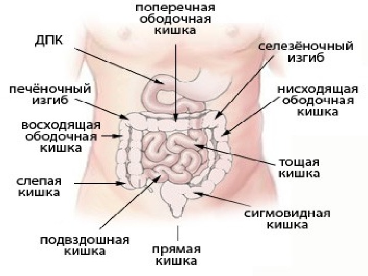
A diagram of the human intestine in pictures with inscriptions in front, will make it possible visually and easily:
- learn everything about the intestines;
- understand where this organ is located;
- study all departments and structural features of the intestines.
What is the intestine, anatomy
In a healthy person, the intestinal walls are lined with intestinal villi and their microvilli, which help to increase the absorbing surface of the nutrients we ingest and bring them into the bloodstream to reach the organs according to their needs. Therefore, in trauma, absorption does not occur optimally and a deficiency occurs. The most common is iron, which causes what can become chronic, as indicated in the guide. When there is anemia and there is no iron recovery after supplementation, we have to think of a different origin, and celiac disease is often that origin.
The intestines are one of the most important human organs.
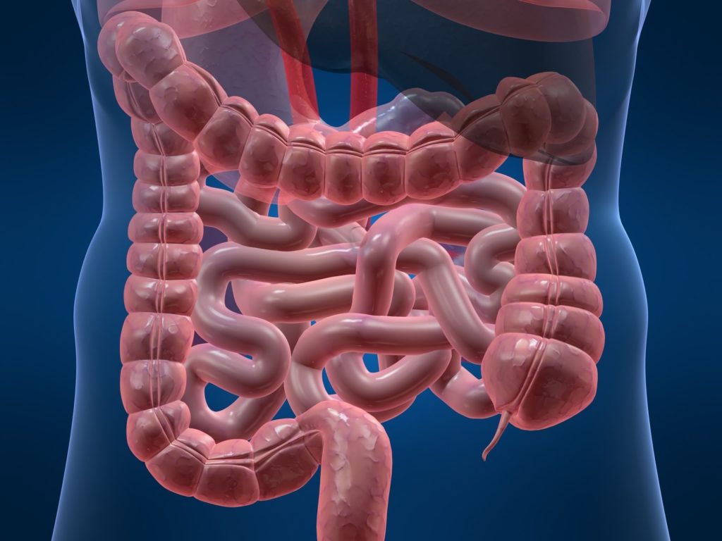
The intestine is the human digestive and excretory organ. The volumetric image clearly demonstrates the structure diagram: what the human intestine consists of and what it looks like.
It is located in the abdominal space and consists of two segments: thin and thick.
There are two sources of its blood supply:
In people with celiac disease, the intestinal villi are the first to suffer damage caused by our body's immune response. This is why it is so important, because it will determine if there is trauma in the intestinal villi and what the extent of the trauma is. The extent of the injury is determined by the creation of the British pathologist of the same name.
Parameters and diseases of the intestine
The structure of intestinal villi is indeed complex despite its minimal size, but the function they perform is of paramount importance for optimal health. The cells of the immune system located in the intestine are called intraepithelial lymphocytes. When these cells get confused and react inappropriately, they can cause inflammatory disorders in both the small intestine and the colon. Among these diseases are Crohn's disease and celiac disease.
- Thin- we supply blood from the superior mesenteric artery and celiac trunk
- Thick- from the superior and inferior mesenteric artery.
The starting point of the intestinal structure is the pylorus of the stomach, and it ends with the anus.
Being in constant activity, the length of the intestine in a living person is about four meters; after death, the muscles relax and provoke its increase in size up to eight meters.
The structure of the rectum of the large intestine
Celiac disease, as explained in the manual, is a disease in which the presence of changes in populations of intestinal intraepithelial lymphocytes is better documented. These changes lead to an increase in the total lymphocyte count, and this number is used to determine if there is an injury and how severe it is, according to Marsh's classification. Other structural elements of the intestinal villi are also being studied.
Marsh, as the manual says. Marsh's classification shows three degrees of injury: infiltrative class 1, infiltrative-hyperplastic class 2, destructive class 3 flat, from the least to the most severe. Therefore, when we talk about the degree of celiac disease, we are referring only and exclusively to the degree of intestinal damage. This means that there is more celiac disease without celiac disease than others, and the least amount of gluten causes internal damage at the gut level, whether or not external symptoms are present.
The intestine grows with the human body, changing its size, diameter, thickness.
So in a newborn child, its length is about three meters, and the period of intensive growth is the age from five months to five years, when the child passes from breastfeeding to the common "table" and larger portions.
Marsh's scores in their classification have the following characteristics. The degree of damage The structure of the villi does not change, but the number of intraepithelial lymphocytes exceeds 25%. It is the most common celiac disease in adults, but it does not always indicate celiac disease, but it can also be caused by other diseases.
Intestinal villi: biopsy
Degree of damage The structure of the villi is normal, but contains hyperplastic crypts, as well as intraepithelial lymphocytosis in greater numbers. It is subdivided to distinguish between the degree of villus atrophy in partial, intermediate and total. It is one of the most important tests for diagnosing celiac disease. In fact, when there are diagnostic doubts, the bowel biopsy usually has the final say. Experts recommend having a bowel biopsy to check the extent of bowel damage regardless of whether celiac disease is confirmed.
The intestine performs the following functions in the human body:
- Provides the intake of hydrochloric acid in the stomach for the primary processing of food;
- Actively participates in the digestive process, splitting the eaten food into separate components and taking from them the trace elements necessary for the body, water;
- Forms and removes feces from the body;
- It has an important effect on the hormonal and immune systems of a person;
The small intestine and its functions
But perhaps it is more correct to treat not the effect, but the cause?
Intestine biopsy, as we explained in the manual, when removing a small piece of the most superficial region of the intestine to detect and analyze possible changes in its shape and in its cells. Today, this is the definitive test for diagnosis, although sometimes other tests can confirm this. Therefore, all experts recommend doing it, even if it is confirmed that celiac disease identifies the degree of damage to the intestine of celiac disease.
Although a bowel biopsy is an invasive test, it is not painful as the patient is sedated. The test, which is part of the histological examination of celiac disease, involves inserting an endoscope, a flexible tube, through the patient's mouth and collecting some samples for analysis. Experts recommend that three samples be taken specifically from the second or third part of the duodenum, the part of the small intestine that connects the stomach to the jejunum, and a sample from the bulb, the first section of the duodenum.
The diagram clearly shows the location of the small intestine between the stomach and the large intestine.
The small intestine is responsible for the digestive process, and it is named so because of its relatively smaller diameter and thinner walls, in contrast to the large intestine. But in its size it is not inferior to any organ of the digestive tract, capturing almost the entire lower space of the peritoneum and partly of the small pelvis.
It measures 7 meters It has 3 regions Place of absorption of digestive products Simple columnar epithelium with goblet cells and microvilli. Cubic cells: unicellular glands. Payer Peyer's: lymphoid nodules. extravasated leukocytes. Mucous membrane, which helps to neutralize acidic chemicals. Brunner's iron: give off an alkaline liquid.
Muscular self: Auerbach's plexus: internal neuronal innervation. Small intestine cells. Cubic cells Monocellular glands The duodenum contains the lowest number of goblet cells and increases to the ileum. mucus component. . a protective layer covering the light.
General work enzymes of the small intestine, gallbladder and the pancreas, promotes the breakdown of food into single components. Here, the absorption of vitamins, nutrients, and active components of most medicines, necessary for the human body, takes place.
Regenerative cells These are stem cells that multiply widely to fill the epithelium of crypts and villi. Unique oval core located at the base . Tumor of the stomach Performed by the stomach. The main irritants assigned by the duodenum: ◦ High acidity ◦ Hypertension ◦ Presence of protein digestion products ◦ Fat digestion product. Regulation of gastric emptying. The duodenum controls gastric emptying through the enterogastric reflex.
. and a pyloric pump stimulator. The spread and presence of certain foods in the stomach stimulates the secretion of gastrin. Gastrin is an enhancer of the secretion of highly acidic gastric juice. Regulation of gastric emptying Gastric factors An increase in the volume of food in the stomach stimulates its emptying: the stretching of the stomach wall awakens myeral reflexes This wall strongly stimulates the activity of the pyloric pump and at the same time slightly inhibits the pylorus.
In addition to the digestive and absorption function, it is responsible for:
- moving food masses further along the intestines;
- strengthening the immune system;
- hormonal secretion.
This segment is subdivided according to the structure scheme into three sections: duodenum 12, jejunum, ileum.
Duodenal
Structure and function
The degree of acidity of the duodenal chemo. The presence of irritation of the mucous membrane of the duodenum. Regulation of gastric emptying Duodenal factors In the duodenum, foods provoke reflexes that reduce gastric emptying.
The factors that can excite enterogastric reflexes are: the presence of certain degradation products in the chyme. . The stimuli for these hormones are mainly fats that enter the duodenum. Regulation of gastric emptying Hormones released from the upper intestine inhibit gastric emptying. More potent hormone: cholecystokinin Another may be a secretor and inhibitor of the liver of the stomach.
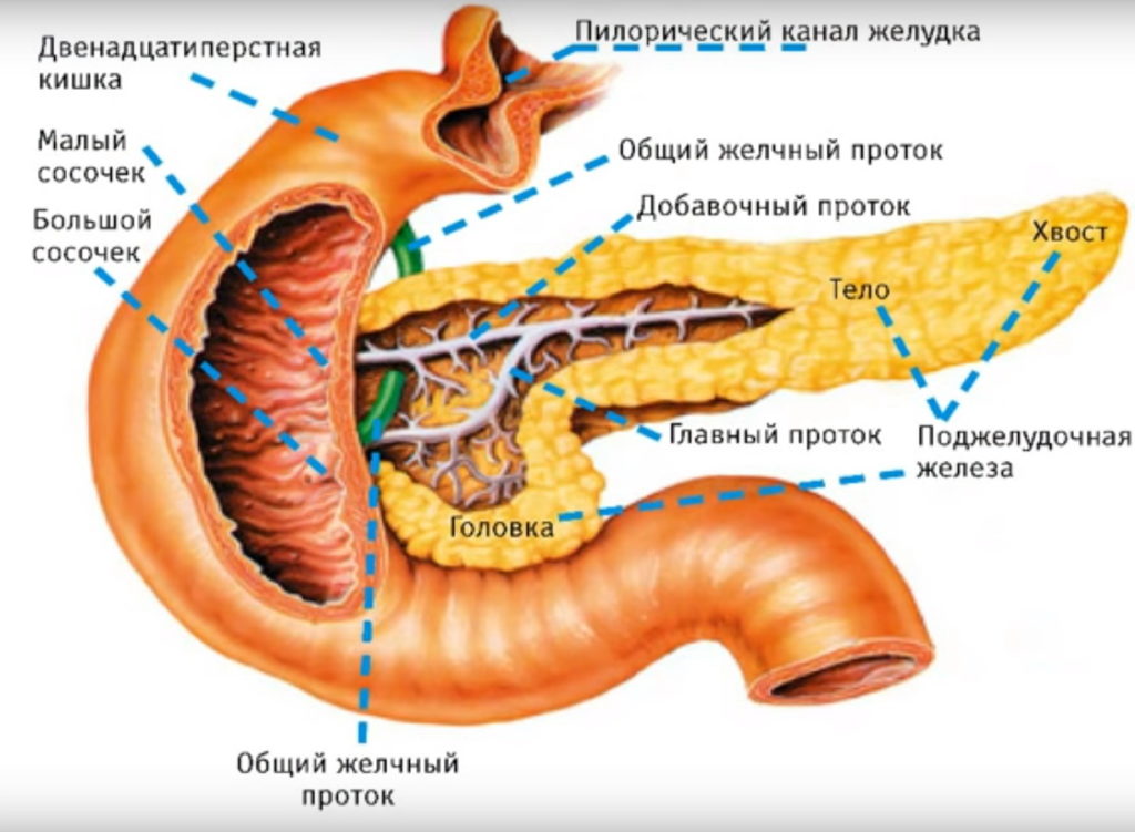
Opens the beginning of the structure of the small intestine - the duodenum, stretching behind the pylorus of the stomach, fitting the head and partly the body of the pancreas, thereby forming the shape of a "horseshoe" or half-ring and pours into the jejunum.
Consists of four parts:
- Top;
- Downward;
- Lower;
- Ascending.
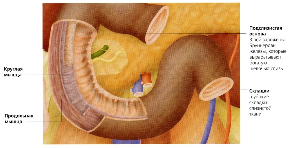
In the middle of the descending part, at the end of the longitudinal fold of the mucous layer, there is the Vater nipple, which includes the sphincter of Oddi. The flow of bile and digestive juice into the duodenum is regulated by this sphincter, and it is also responsible for excluding the penetration of its contents into the bile and pancreatic ducts.
This promotes the formation of pancreatic juice. Dissolves nucleoproteins and solubilizes collagen. Regulates the opening and closing of the pylor. Disaccharide hydrolysis: sucrose 6 glucose fructose. This facilitates the conversion of pepsinogen to pepsin. The small intestine is the part of the digestive tract that begins after the stomach and ends in the cecum of the large intestine. It is divided into three sections: the duodenum, the jejunum, and the ileum. The intestinal wall consists of three layers, the first is the mucous membrane of the musculature, which unites the outer surface and separates the mucous membrane from the submucosa; The second layer is a sheet, which is an intermediate layer and consists of connective tissue.
Skinny
The anatomical structure separating the jejunum and ileum is weak, but there is still a difference. The ilium is relatively skinny, larger in diameter and has thicker walls. She was named skinny due to the lack of content in it during the autopsy. The jejunum can be up to 180 cm long and is longer in men than in women.
This layer contains blood vessels, lymph vessels, nerves, smooth muscle tissue, connective tissue, and white blood cells. In the epithelial cells of the intestine, intestinal hairs are located, which are elongations of approximately 5-5 mm, the function of which is to increase the contact surface area and therefore increase the absorption surface. These villi are wider in the duodenum than in the rest of the intestine. The presence of hair is of paramount importance for optimal function of the small intestine.
The absorption surface is made even larger by the small brushes that cover the intestinal hair, called microvilli. Microvillins are covered with membranes that protect them from proteolytic and mucolytic agents. Any toxic substance that alters the morphological structure of hair and microfiber will affect absorption and therefore lead to possible malnutrition by decreasing the absorption of proteins, essential minerals and other nutrients.
Iliac
The description of the diagram of the structure of the lower part of the small intestine (diagram above) is as follows: following the jejunum, the ileum is connected to the upper part of the large intestine by means of the Bauhinia valve; placed on the bottom right abdominal cavity... Above are the distinctive properties of the ileum from the jejunum. But general characteristic these parts of the human intestine is a clear expression of the mesentery.
Himph, which is created in the stomach, a bolus mixed with hydrochloric acid from peristaltic movements, mixed with bile and pancreatic secretions, so as not to break the layers of the small intestine and not get into the duodenum. The phenomenon of digestion and absorption largely depends on the contact of food with the intestinal walls, so that the more it is and on a wider surface, the better the digestion and absorption of food will be.
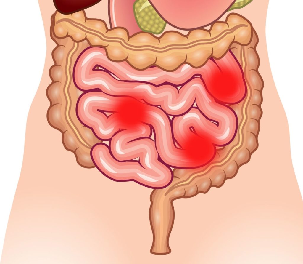
Symptoms of Small Bowel Problems
This gives us one of the most important morphological characteristics of the small intestine, which are numerous folds that enhance the absorption surface, for example: Intestinal villi. Microvillins in epithelial cells. Anatomically, the small intestine is a two to three meter long tube extending from the pylorus to the ileocecal valve. It is found in the central and lower abdomen. It is mobile, except for the duodenum, which is retroperitoneal and fixed.
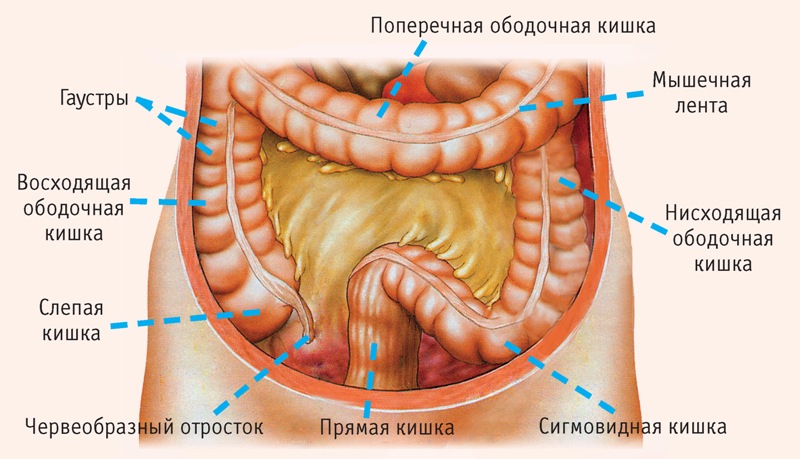
Colon
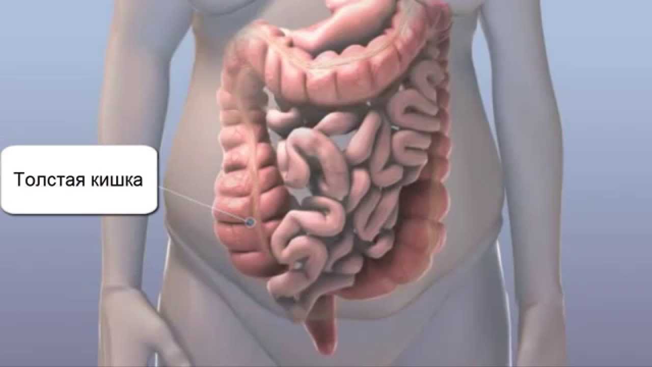
The lower and last segment of the gastrointestinal tract and intestines is the large intestine, which is responsible for water absorption and formation feces from chyme. The figure shows the layout of this part of the intestine: in the abdominal space and the cavity of the small pelvis.
The structural features of the colon wall are contained in the mucous layer, which protects from the inside from the negative influence of digestive enzymes, mechanical injury by solid particles of feces and simplifies its movement to the exit. Human desires are not subject to the work of the intestinal musculature, it is absolutely independent and not controlled by humans.
The structure of the intestine starts from the ileocecal valve and ends with the anus. Like the small intestine, it has three anatomical segments with the following names: blind, colon and rectum.
Blind
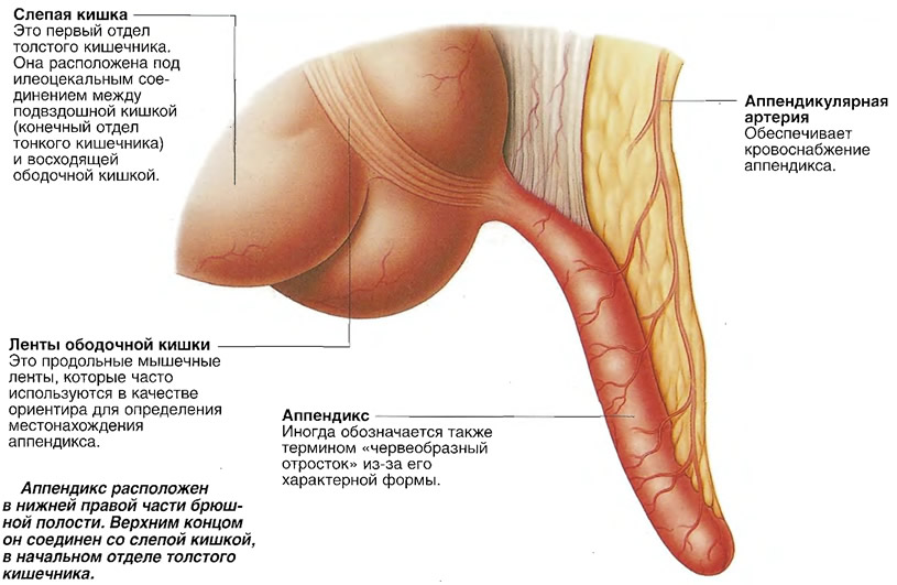
The original section of the human large intestine, located according to the scheme in the right iliac fossa, framed from everywhere by the peritoneum, is arranged in the form of a sac.
From the back wall of the cecum, its appendage is secreted, nothing more than the appendix, a tubular process about ten cm in size and one cm in diameter, performing secondary functions necessary for the human body: it produces amylase, lipase and hormones involved in the work of intestinal sphincters and peristalsis.
Colonic
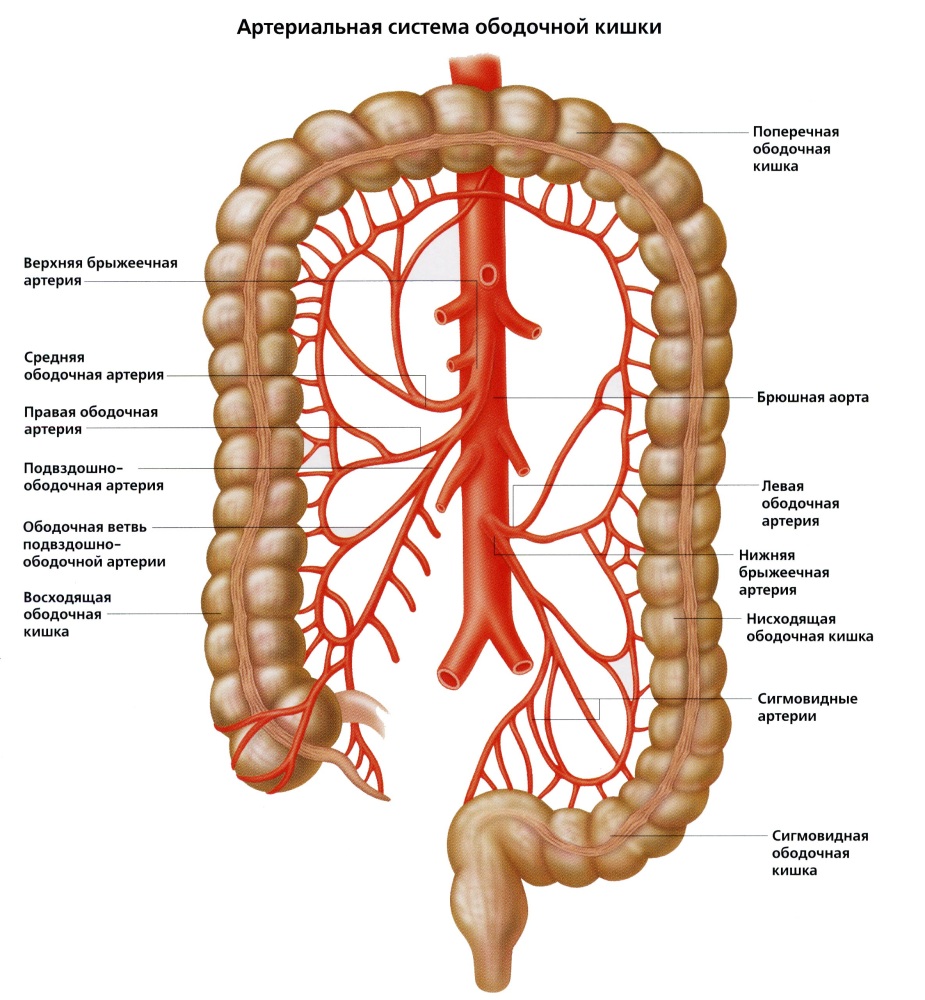
The main part of the large intestine is located according to the structure between the cecum and rectum.
At the junction with the blind, an ascending cecum sphincter is placed. The colon is subdivided into the following segments:
- Ascending;
- Transverse;
- Falling;
- Sigmoid.
Here, water and electrolytes are absorbed in large quantities, as well as the transformation of liquid chyme into hardened, shaped feces.
Straight
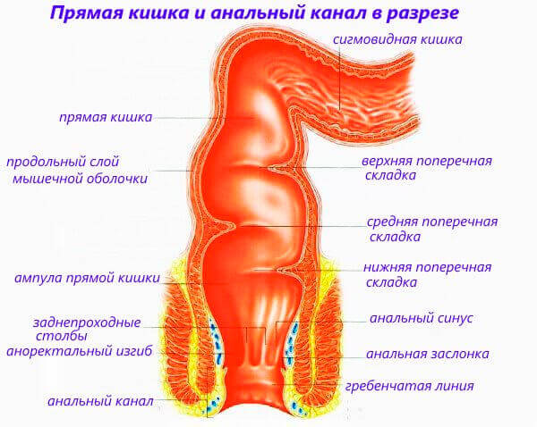
Placed within the small pelvis and free of volvulus - the rectum completes the structure of the large intestine, starting from sigmoid colon(level of the third sacral vertebra) and ending with the anus (perineal region). Here feces accumulate, controlled by two sphincters of the anus (internal and external). The sectional diagram of the intestine demonstrates its division into two sections: a narrow (anal canal) and a wide (ampullary).
Parameters and diseases of the intestine
| Intestine and its departments | Parameters (cm) | Diseases | |
|---|---|---|---|
| Length | Diameter | ||
| Small intestine | 350-400 | Proximal 4-6, distal 2-3. | Obstruction, ciliakia, enteritis, colitis, Meckel's diverticulum. |
| Duodenal | 21-25 | 42859 | Duodenitis, ulcer |
| Skinny | 90-180 | Yunit, ulcer, | |
| Iliac | 130-260 | 2.7 | Crohn's disease, terminal ileitis. |
| Colon | 150-200 | 42951 | Atony, cancer, inflammation. |
| Blind | 3-8,5 | 7 | Cancer, appendicitis. |
| Colonic | About 150 | 5 -8 | IBS, agangliosis, regional enteritis, colitis, constipation and diarrhea, intestinal intussusception. |
| Straight | 14-18 | 4-7,5 | Cancer, hemorrhoids. |
The general state of human health depends on the normal functioning of the intestines. This organ is susceptible to various disorders, disorders and inflammations, often due to the fault of a person with errors in nutrition. Serious pathologies are also no exception - to prevent their development and exacerbation, it is recommended to monitor your health, undergo unscheduled examinations, and lead a healthy lifestyle.
Located between the stomach and the large intestine. By the name you can judge the thickness of its walls. They are thinner than those of the large intestine, and the lumen is narrower.
The small intestine is the longest in the body.
The length of the small intestine can be up to 6 m, this is the longest part. There are three divisions of the small intestine:
- Duodenal. The length of this section is about 12 fingers of the hand folded transversely, that is, about 20 cm. Hence the name. We have a special task - to neutralize gastric acid. Food in the stomach is only partially digested. And then a lump of food, glued together by gastric juice, enters the duodenum, where it is brought into a state that is safe for the rest of the intestine. Therefore, the walls of this section have a special structure that allows the acid to stay calmly inside the intestine without damaging it.
- Skinny and iliac. The length of the jejunum reaches 3 m. It does not have a clear border with the ileum, therefore, they are often considered together, and separated only conditionally. The functions of both departments are similar: to secrete special enzymes that break down food, as well as to assimilate nutrients, etc. The intestine has motility and is also responsible for the regular and correct movement of food through it. The jejunum is approximately at the level of the navel, and the ileum is at the level of the small pelvis. The small intestine consists of several layers: mucous, submucous, muscular and external.
The mucous membrane differs only in the duodenum. There are special glands, and the villi of the epithelium are longer and thicker.
The length and diameter of the lumen increases with the course of life (from childhood to adolescence and adolescence). In children, the small intestine can change depending on its presence, body position, or bowel tone.
Small intestine functions
The small intestine is capable of producing certain hormones.
As mentioned above, the small intestine performs several functions at once. Knowledge of the divisions and basic functions of the small intestine will identify and prevent many:
- Production of a secret for the breakdown of food. The small intestine produces juice, which contains various enzymes that break down carbohydrates into their simplest components. This is the direct and most important task of the small intestine. Only after complete cleavage are nutrients absorbed by the villi of the epithelium.
- The small intestine also performs a motor function. With the help of muscle contractions, unprocessed food debris moves further through the intestines. This is called peristalsis. If it is insufficient, they are formed.
- The ability of the villi to absorb nutrients can be called a separate, absorption function. In some bowel diseases, the absorption of vitamins is impaired, and hypovitaminosis or B12-deficiency anemia begins.
- The small intestine is capable of producing some hormones necessary for the normal functioning of the digestive tract. One of these hormones is secretin. It regulates the work. Its main function is to stimulate the production of pancreatic juice.
- One of the functions of the small intestine is excretory. By breaking down food, the small intestine prepares it for elimination from the body.
- The small intestine is also responsible for immunity. It produces immunoglobulin A, which is responsible for humoral immunity (present in the blood, destroys disease-causing cells). It is also responsible for protecting the mucous membranes, preventing pathogenic microorganisms from penetrating deep into the tissues.
- A separate function is the neutralization of the acidic environment, which is performed by the duodenum. In diseases of the duodenum, acid can enter the intestines, causing irritation and pain.
Diseases of the small intestine
With enteritis, the work of the mucous membrane is disrupted.
Various diseases of the small intestine are most often expressed in stool disorders and. No intestinal disease can do without these symptoms. However, an accurate diagnosis can only be made after an examination. Common diseases:
- Duodenitis. This is a disease of the duodenum. The intestinal mucosa becomes inflamed and cannot fully fulfill its main function - to neutralize acid. The reasons can be abuse, unhealthy diet, including a large number of spicy and irritating foods, as well as various poisoning. In this case, there is abdominal pain, stool disturbance.
- ... As a result of the aggressive effect of acid on the intestinal mucosa, an ulcer is formed, which, even after effective treatment leaves a scar. With an ulcer, patients experience severe, sharp abdominal pains that occur after eating spicy food, strenuous physical exertion, or with a prolonged absence of food.
- small intestine. This disease is quite rare, in about 2% of cases among oncological diseases of all organs of the gastrointestinal tract. Elderly men are more likely to get sick. The reasons can be very different. The provoking factors are smoking, alcohol, in the small intestine, carcinogens in food, ulcers and other bowel diseases that can lead to cancer. Cancer develops slowly, which forces patients to see a doctor only in the last stages, when symptoms are clearly manifested, and the tumor itself has already grown deep into the muscle tissue and has narrowed the intestinal lumen.
- ... With enteritis, the intestinal mucosa changes in such a way that the absorption function is impaired. Enteritis rarely occurs separately. Usually they are accompanied by other diseases of the gastrointestinal tract, for example. The causes of enteritis include malnutrition, alcohol, hereditary diseases, poisoning, infections, allergens.
Diagnostics and treatment
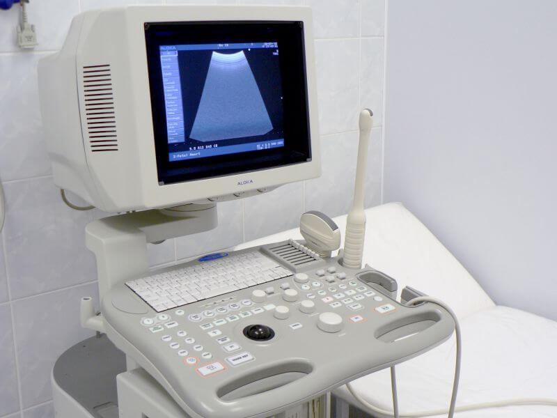
Ultrasound is one of the ways to examine the duodenum.
Examination of the small intestine is fraught with difficulties. The small intestine is positioned in such a way that it can be difficult to reach it with an endoscope. This also explains the insufficient knowledge of diseases of the small intestine. When making a diagnosis, the patient's complaints, blood test results, and other examination results are taken into account.
The most informative diagnostic method is still the video capsule. It is the video capsule endoscope that is able to reach and examine the entire small intestine. For this, a small bowel video capsule is used, which is administered without a tube through the mouth. In other words, you just need to swallow the capsule. The result is displayed on the monitor screen. There is no need to worry about its exit from the body, the capsule is disposable.
In some cases, the capsule can linger in the body, but there has long been a special way to remove it without surgery. If the delay is caused by a serious pathological process, surgery is necessary. Such a diagnosis requires special preparation. Before the procedure, you need to adhere to a special diet, on the day of the procedure, cleanse the intestines and take those that promote the elimination of gases.
In some cases, a biopsy is necessary, when a small area of tissue is taken for a more detailed examination. This is not the easiest procedure, it is carried out using a probe. Various complications are possible, for example, the sticking of the probe in the intestine. Treatment depends on the specific condition. A mandatory point of treatment is always a gentle diet, which reduces the load on the intestines, includes food that does not irritate the mucous membranes and is easily digestible.
You will learn about the study of the small intestine from the video:
Have you noticed a mistake? Highlight it and press Ctrl + Enter to let us know.
Tell your friends! Share this article with your friends on your favorite social network using the social buttons. Thanks!